{"title":"Regional Pulmonary Involvement in Bronchiolitis: Insights From Lung Ultrasound.","authors":"Seyfeddine Zayani, Farah Thabet, Abir Daya, Olfa Betbout, Chokri Chouchane, Slaheddine Chouchane","doi":"10.1002/ppul.71337","DOIUrl":null,"url":null,"abstract":"<p><strong>Background: </strong>Acute bronchiolitis is a leading cause of pediatric hospitalization, with severe cases necessitating ventilatory support. Lung ultrasound (LUS) is emerging as a valuable tool for assessing respiratory conditions, yet its utility in evaluating regional heterogeneity in bronchiolitis remains underexplored.</p><p><strong>Objectives: </strong>This study aimed to assess the regional distribution of pulmonary lesions in infants with bronchiolitis using LUS and explore their association with the need for ventilatory support.</p><p><strong>Methods: </strong>A prospective study of 160 infants with bronchiolitis was conducted at a tertiary care center. LUS was performed within the first 12 h of admission, with pulmonary regions scored based on the Brat scoring system. Patients were categorized into a favorable outcome group and a ventilatory support group, and the severity of regional lung lesions was analyzed.</p><p><strong>Results: </strong>Median age was 65.5 days (IQR 38-118.5; range 11-314). Infants requiring ventilatory support exhibited higher regional LUS scores-particularly in lateral-superior, lateral-inferior, posterior-superior, and posterior-inferior zones (p = 0.001); posterior regions showed the highest prevalence of severe lesions. In multivariable analysis, involvement of specific zones independently predicted ventilatory support, notably right lateral-superior (OR 4.6, 95% CI 2.12-9.86), left lateral-superior (OR 3.7, 95% CI 1.78-7.86), left posterior-superior (OR 2.0, 95% CI 1.23-3.51), and left posterior-inferior (OR 2.1, 95% CI 1.20-3.71).</p><p><strong>Conclusions: </strong>Our findings highlight the heterogeneous distribution of pulmonary involvement in bronchiolitis and underscore the potential role of LUS in severity stratification. However, the study's single-center design necessitates cautious interpretation, with further research needed to validate these results and expand the clinical application of LUS in bronchiolitis management.</p>","PeriodicalId":19932,"journal":{"name":"Pediatric Pulmonology","volume":"60 10","pages":"e71337"},"PeriodicalIF":2.3000,"publicationDate":"2025-10-01","publicationTypes":"Journal Article","fieldsOfStudy":null,"isOpenAccess":false,"openAccessPdf":"https://www.ncbi.nlm.nih.gov/pmc/articles/PMC12522026/pdf/","citationCount":"0","resultStr":null,"platform":"Semanticscholar","paperid":null,"PeriodicalName":"Pediatric Pulmonology","FirstCategoryId":"3","ListUrlMain":"https://doi.org/10.1002/ppul.71337","RegionNum":3,"RegionCategory":"医学","ArticlePicture":[],"TitleCN":null,"AbstractTextCN":null,"PMCID":null,"EPubDate":"","PubModel":"","JCR":"Q1","JCRName":"PEDIATRICS","Score":null,"Total":0}
引用次数: 0
Abstract
Background: Acute bronchiolitis is a leading cause of pediatric hospitalization, with severe cases necessitating ventilatory support. Lung ultrasound (LUS) is emerging as a valuable tool for assessing respiratory conditions, yet its utility in evaluating regional heterogeneity in bronchiolitis remains underexplored.
Objectives: This study aimed to assess the regional distribution of pulmonary lesions in infants with bronchiolitis using LUS and explore their association with the need for ventilatory support.
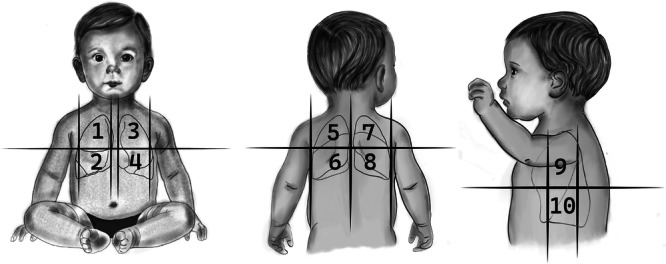
Methods: A prospective study of 160 infants with bronchiolitis was conducted at a tertiary care center. LUS was performed within the first 12 h of admission, with pulmonary regions scored based on the Brat scoring system. Patients were categorized into a favorable outcome group and a ventilatory support group, and the severity of regional lung lesions was analyzed.
Results: Median age was 65.5 days (IQR 38-118.5; range 11-314). Infants requiring ventilatory support exhibited higher regional LUS scores-particularly in lateral-superior, lateral-inferior, posterior-superior, and posterior-inferior zones (p = 0.001); posterior regions showed the highest prevalence of severe lesions. In multivariable analysis, involvement of specific zones independently predicted ventilatory support, notably right lateral-superior (OR 4.6, 95% CI 2.12-9.86), left lateral-superior (OR 3.7, 95% CI 1.78-7.86), left posterior-superior (OR 2.0, 95% CI 1.23-3.51), and left posterior-inferior (OR 2.1, 95% CI 1.20-3.71).
Conclusions: Our findings highlight the heterogeneous distribution of pulmonary involvement in bronchiolitis and underscore the potential role of LUS in severity stratification. However, the study's single-center design necessitates cautious interpretation, with further research needed to validate these results and expand the clinical application of LUS in bronchiolitis management.
背景:急性细支气管炎是儿童住院的主要原因,严重者需要呼吸机支持。肺超声(LUS)正在成为评估呼吸系统疾病的一种有价值的工具,但其在评估毛细支气管炎区域异质性方面的应用仍未得到充分探讨。目的:本研究旨在评估使用LUS的毛细支气管炎婴儿肺部病变的区域分布,并探讨其与呼吸支持需求的关系。方法:在三级保健中心对160例毛细支气管炎婴儿进行前瞻性研究。在入院前12小时内进行LUS,根据Brat评分系统对肺区域进行评分。将患者分为预后良好组和通气支持组,并分析局部肺病变的严重程度。结果:中位年龄为65.5天(IQR 38-118.5;范围11-314)。需要呼吸支持的婴儿表现出更高的区域LUS评分,特别是在侧上区、侧下区、后上区和后下区(p = 0.001);后区显示严重病变的发生率最高。在多变量分析中,特定区域的介入独立预测了通气支持,特别是右侧上方(OR 4.6, 95% CI 2.12-9.86)、左侧上方(OR 3.7, 95% CI 1.78-7.86)、左后上方(OR 2.0, 95% CI 1.23-3.51)和左后下方(OR 2.1, 95% CI 1.20-3.71)。结论:我们的研究结果强调了细支气管炎肺部受累的异质性分布,并强调了LUS在严重程度分层中的潜在作用。然而,该研究的单中心设计需要谨慎的解释,需要进一步的研究来验证这些结果并扩大LUS在毛细支气管炎治疗中的临床应用。
期刊介绍:
Pediatric Pulmonology (PPUL) is the foremost global journal studying the respiratory system in disease and in health as it develops from intrauterine life though adolescence to adulthood. Combining explicit and informative analysis of clinical as well as basic scientific research, PPUL provides a look at the many facets of respiratory system disorders in infants and children, ranging from pathological anatomy, developmental issues, and pathophysiology to infectious disease, asthma, cystic fibrosis, and airborne toxins. Focused attention is given to the reporting of diagnostic and therapeutic methods for neonates, preschool children, and adolescents, the enduring effects of childhood respiratory diseases, and newly described infectious diseases.
PPUL concentrates on subject matters of crucial interest to specialists preparing for the Pediatric Subspecialty Examinations in the United States and other countries. With its attentive coverage and extensive clinical data, this journal is a principle source for pediatricians in practice and in training and a must have for all pediatric pulmonologists.
 求助内容:
求助内容: 应助结果提醒方式:


