{"title":"Complications of CT-Guided Percutaneous Lung Biopsy: A 1-Year Single-Center Experience in Iran.","authors":"Mohammad Reza Sasani, Majid Paknahad","doi":"10.47176/mjiri.39.102","DOIUrl":null,"url":null,"abstract":"<p><strong>Background: </strong>Despite observing all precautions, complications are not uncommon during transthoracic needle biopsy (TTNB). We aimed to evaluate the complications associated with TTNB in patients with lung masses from 1 center in southern Iran.</p><p><strong>Methods: </strong>In this retrospective cohort study, data on complication rates, types, and potential risk factors from 87 biopsies were collected. Complications were assessed through immediate post-biopsy computed tomography (CT) scans and follow-up chest X-rays, and their correlations were evaluated with patient demographics, lesion characteristics, and procedural factors. Chi-square and Wilcoxon rank-sum tests were used for univariable analysis, and multivariable binary logistic regression analyses were conducted to control for potential confounders.</p><p><strong>Results: </strong>The overall complication rate was 37.9% (95% CI, 27.6%-48.3%), with pneumothorax being the most common, occurring in 26.4% (95% CI, 17.3%-35.6%) of cases, followed by perilesional hemorrhage (17.2%) (95% CI, 10.3%-25.3%), hemoptysis (3.3%), and pleural effusion (1.1%). All pneumothorax cases were identified via immediate post-biopsy CT, and only 1 patient required chest tube insertion. No significant correlations were found between age, sex, presence of emphysema, lesion size, location, and depth, or needle path and the incidence of pneumothorax. However, a significantly higher perilesional hemorrhage incidence was observed for smaller lesion size (26 mm [interquartile range, IQR], 13,40 vs 43 mm [IQR, 24,73]; <i>P</i> = 0.019), deeper lesion (10 mm [IQR, 0.17] vs 0 mm [IQR, 0.10]; <i>P</i> = 0.041), and longer needle path (17 mm [IQR, 9.29] vs 0 mm [IQR, 0.7]; <i>P</i> < 0.001). Furthermore, 47.8% of pneumothorax cases identified on postbiopsy CT showed no signs on follow-up chest X-ray 3 hours later.</p><p><strong>Conclusion: </strong>TTNB is generally safe, with a manageable complication profile. Early detection and appropriate follow-up are crucial, particularly for pneumothorax, which often resolves spontaneously. The findings underscore the importance of considering lesion characteristics to minimize complications during biopsy procedures.</p>","PeriodicalId":18361,"journal":{"name":"Medical Journal of the Islamic Republic of Iran","volume":"39 ","pages":"102"},"PeriodicalIF":0.0000,"publicationDate":"2025-08-04","publicationTypes":"Journal Article","fieldsOfStudy":null,"isOpenAccess":false,"openAccessPdf":"https://www.ncbi.nlm.nih.gov/pmc/articles/PMC12516420/pdf/","citationCount":"0","resultStr":null,"platform":"Semanticscholar","paperid":null,"PeriodicalName":"Medical Journal of the Islamic Republic of Iran","FirstCategoryId":"1085","ListUrlMain":"https://doi.org/10.47176/mjiri.39.102","RegionNum":0,"RegionCategory":null,"ArticlePicture":[],"TitleCN":null,"AbstractTextCN":null,"PMCID":null,"EPubDate":"2025/1/1 0:00:00","PubModel":"eCollection","JCR":"Q2","JCRName":"Medicine","Score":null,"Total":0}
引用次数: 0
Abstract
Background: Despite observing all precautions, complications are not uncommon during transthoracic needle biopsy (TTNB). We aimed to evaluate the complications associated with TTNB in patients with lung masses from 1 center in southern Iran.
Methods: In this retrospective cohort study, data on complication rates, types, and potential risk factors from 87 biopsies were collected. Complications were assessed through immediate post-biopsy computed tomography (CT) scans and follow-up chest X-rays, and their correlations were evaluated with patient demographics, lesion characteristics, and procedural factors. Chi-square and Wilcoxon rank-sum tests were used for univariable analysis, and multivariable binary logistic regression analyses were conducted to control for potential confounders.
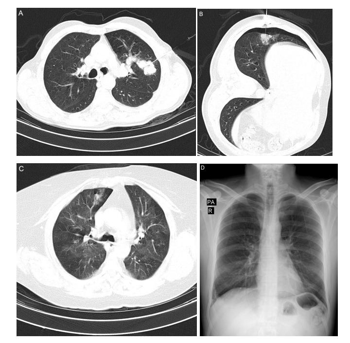
Results: The overall complication rate was 37.9% (95% CI, 27.6%-48.3%), with pneumothorax being the most common, occurring in 26.4% (95% CI, 17.3%-35.6%) of cases, followed by perilesional hemorrhage (17.2%) (95% CI, 10.3%-25.3%), hemoptysis (3.3%), and pleural effusion (1.1%). All pneumothorax cases were identified via immediate post-biopsy CT, and only 1 patient required chest tube insertion. No significant correlations were found between age, sex, presence of emphysema, lesion size, location, and depth, or needle path and the incidence of pneumothorax. However, a significantly higher perilesional hemorrhage incidence was observed for smaller lesion size (26 mm [interquartile range, IQR], 13,40 vs 43 mm [IQR, 24,73]; P = 0.019), deeper lesion (10 mm [IQR, 0.17] vs 0 mm [IQR, 0.10]; P = 0.041), and longer needle path (17 mm [IQR, 9.29] vs 0 mm [IQR, 0.7]; P < 0.001). Furthermore, 47.8% of pneumothorax cases identified on postbiopsy CT showed no signs on follow-up chest X-ray 3 hours later.
Conclusion: TTNB is generally safe, with a manageable complication profile. Early detection and appropriate follow-up are crucial, particularly for pneumothorax, which often resolves spontaneously. The findings underscore the importance of considering lesion characteristics to minimize complications during biopsy procedures.
背景:尽管观察了所有的预防措施,并发症在经胸穿刺活检(TTNB)中并不罕见。我们的目的是评估来自伊朗南部一个中心的肺肿块患者与TTNB相关的并发症。方法:在回顾性队列研究中,收集了87例活检的并发症发生率、类型和潜在危险因素。通过活检后立即进行计算机断层扫描(CT)和随访胸部x光片评估并发症,并评估其与患者人口统计学、病变特征和手术因素的相关性。单变量分析采用卡方检验和Wilcoxon秩和检验,多变量二元逻辑回归分析控制潜在混杂因素。结果:总并发症发生率为37.9% (95% CI, 27.6% ~ 48.3%),以气胸最为常见,发生率为26.4% (95% CI, 17.3% ~ 35.6%),其次为病灶周围出血(17.2%)(95% CI, 10.3% ~ 25.3%)、咯血(3.3%)、胸腔积液(1.1%)。所有气胸病例均通过活检后立即CT确诊,仅有1例患者需要插入胸管。年龄、性别、是否有肺气肿、病灶大小、位置、深度或穿刺路径与气胸发生率无显著相关性。然而,病灶较小(26 mm[四分位间距,IQR], 13,40 vs 43 mm [IQR, 24,73], P = 0.019),病灶较深(10 mm [IQR, 0.17] vs 0 mm [IQR, 0.10], P = 0.041),针径较长(17 mm [IQR, 9.29] vs 0 mm [IQR, 0.7], P < 0.001),病灶周围出血发生率明显较高。此外,47.8%的活检后CT发现的气胸病例在3小时后的随访胸片中没有任何迹象。结论:TTNB总体上是安全的,并发症可控。早期发现和适当的随访是至关重要的,特别是对于气胸,它往往是自发解决的。研究结果强调了在活检过程中考虑病变特征以减少并发症的重要性。

 求助内容:
求助内容: 应助结果提醒方式:


