Gertruda Evaristo, Namrata Setia, Peng Wang, Peter Pytel, Lindsay Alpert
{"title":"Low-grade NTRK-rearranged spindle cell neoplasm presenting as a colonic polyp and managed by polypectomy: a rare case report and literature review.","authors":"Gertruda Evaristo, Namrata Setia, Peng Wang, Peter Pytel, Lindsay Alpert","doi":"10.1186/s13000-025-01713-3","DOIUrl":null,"url":null,"abstract":"<p><strong>Background: </strong>NTRK-rearranged spindle cell neoplasms constitute a novel, heterogeneous group of mesenchymal neoplasms originally described predominantly in soft tissue locations. They are commonly characterized by co-expression of S100 and CD34 immunostains and presence of NTRK fusions. While exceedingly rare, there are increasing reports of this lesion involving the gastrointestinal tract, presenting predominantly as large masses of the stomach, small bowel and colorectum.</p><p><strong>Case presentation: </strong>We present a case of a 37-year-old male who on colonoscopy was found to have a one cm polyp of the sigmoid colon which was removed by hot snare polypectomy. Histologic examination revealed haphazardly arranged bland spindle cells with diffuse CD34 and S100 co-expression. A targeted Next-Generation RNA Fusion Assay identified a TPR::NTRK1 fusion, confirming the diagnosis of low-grade NTRK-rearranged spindle cell neoplasm. The mucosal and deep margins were free of tumor. In contrast to the previously reported cases, the patient was managed with polypectomy and active surveillance, and remained disease-free at 14 months follow up.</p><p><strong>Conclusion: </strong>This case contributes to the limited body of literature on gastrointestinal low-grade NTRK-rearranged spindle cell neoplasms and raises the possibility of endoscopic treatment consideration for carefully selected patients.</p>","PeriodicalId":11237,"journal":{"name":"Diagnostic Pathology","volume":"20 1","pages":"111"},"PeriodicalIF":2.3000,"publicationDate":"2025-10-14","publicationTypes":"Journal Article","fieldsOfStudy":null,"isOpenAccess":false,"openAccessPdf":"https://www.ncbi.nlm.nih.gov/pmc/articles/PMC12522387/pdf/","citationCount":"0","resultStr":null,"platform":"Semanticscholar","paperid":null,"PeriodicalName":"Diagnostic Pathology","FirstCategoryId":"3","ListUrlMain":"https://doi.org/10.1186/s13000-025-01713-3","RegionNum":3,"RegionCategory":"医学","ArticlePicture":[],"TitleCN":null,"AbstractTextCN":null,"PMCID":null,"EPubDate":"","PubModel":"","JCR":"Q2","JCRName":"PATHOLOGY","Score":null,"Total":0}
引用次数: 0
Abstract
Background: NTRK-rearranged spindle cell neoplasms constitute a novel, heterogeneous group of mesenchymal neoplasms originally described predominantly in soft tissue locations. They are commonly characterized by co-expression of S100 and CD34 immunostains and presence of NTRK fusions. While exceedingly rare, there are increasing reports of this lesion involving the gastrointestinal tract, presenting predominantly as large masses of the stomach, small bowel and colorectum.
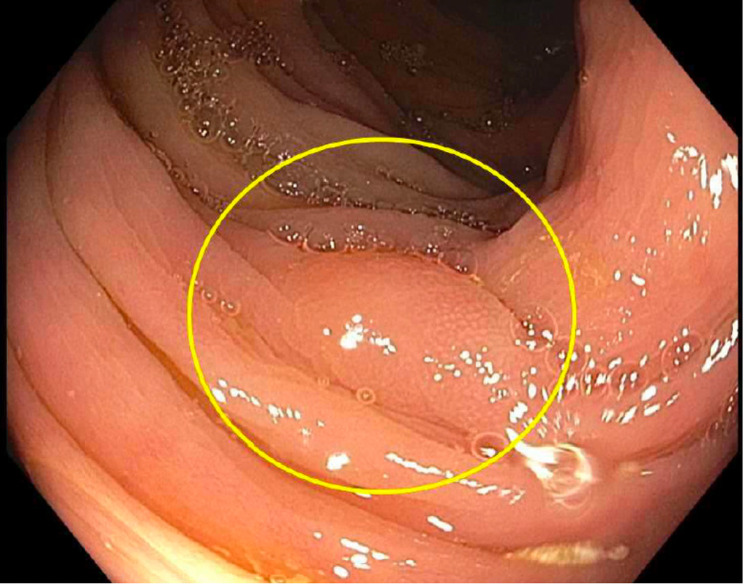
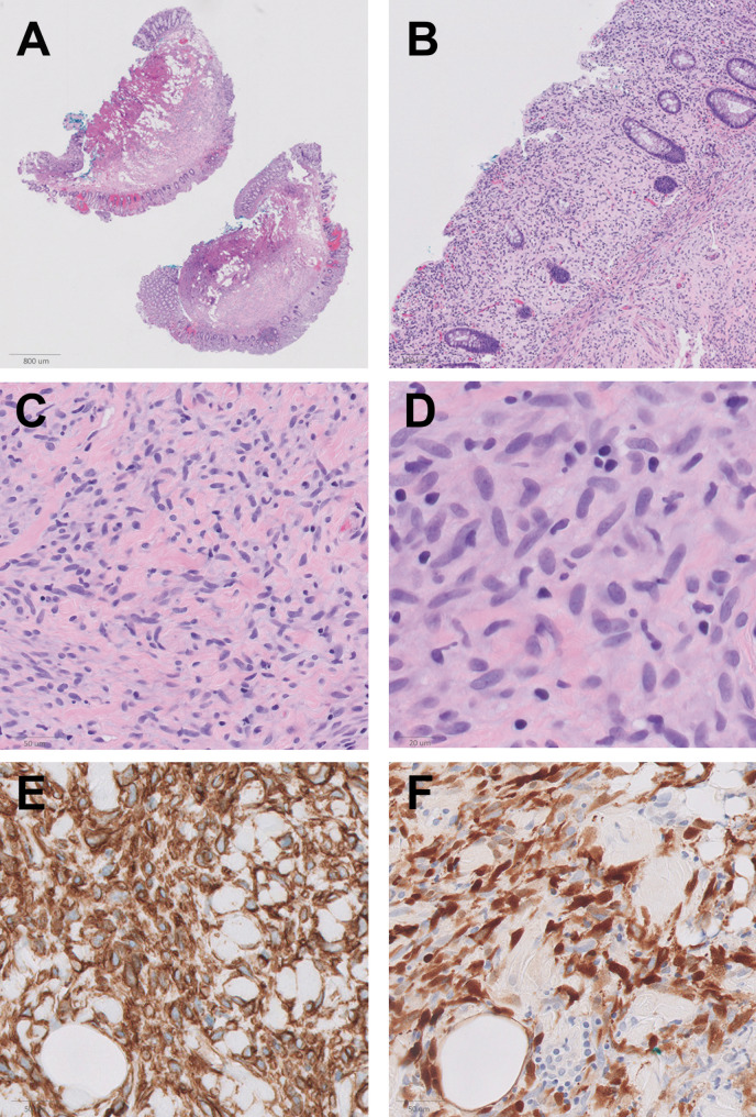
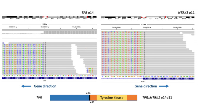
Case presentation: We present a case of a 37-year-old male who on colonoscopy was found to have a one cm polyp of the sigmoid colon which was removed by hot snare polypectomy. Histologic examination revealed haphazardly arranged bland spindle cells with diffuse CD34 and S100 co-expression. A targeted Next-Generation RNA Fusion Assay identified a TPR::NTRK1 fusion, confirming the diagnosis of low-grade NTRK-rearranged spindle cell neoplasm. The mucosal and deep margins were free of tumor. In contrast to the previously reported cases, the patient was managed with polypectomy and active surveillance, and remained disease-free at 14 months follow up.
Conclusion: This case contributes to the limited body of literature on gastrointestinal low-grade NTRK-rearranged spindle cell neoplasms and raises the possibility of endoscopic treatment consideration for carefully selected patients.
期刊介绍:
Diagnostic Pathology is an open access, peer-reviewed, online journal that considers research in surgical and clinical pathology, immunology, and biology, with a special focus on cutting-edge approaches in diagnostic pathology and tissue-based therapy. The journal covers all aspects of surgical pathology, including classic diagnostic pathology, prognosis-related diagnosis (tumor stages, prognosis markers, such as MIB-percentage, hormone receptors, etc.), and therapy-related findings. The journal also focuses on the technological aspects of pathology, including molecular biology techniques, morphometry aspects (stereology, DNA analysis, syntactic structure analysis), communication aspects (telecommunication, virtual microscopy, virtual pathology institutions, etc.), and electronic education and quality assurance (for example interactive publication, on-line references with automated updating, etc.).

 求助内容:
求助内容: 应助结果提醒方式:


