Exploring the utility of cellular indices in the diagnosis of ulcerative colitis

Julio César Moreno Alfonso , Sharom Barbosa-Velásquez , Carlos Delgado-Miguel , Ada Molina Caballero , Sara Hernández-Martín , Alberto Pérez Martínez , María Concepción Yárnoz Irazábal
{"title":"Exploring the utility of cellular indices in the diagnosis of ulcerative colitis","authors":"Julio César Moreno Alfonso ,&nbsp;Sharom Barbosa-Velásquez ,&nbsp;Carlos Delgado-Miguel ,&nbsp;Ada Molina Caballero ,&nbsp;Sara Hernández-Martín ,&nbsp;Alberto Pérez Martínez ,&nbsp;María Concepción Yárnoz Irazábal","doi":"10.1016/j.gastre.2025.502349","DOIUrl":null,"url":null,"abstract":"<div><h3>Aim</h3><div>To describe the usefulness of cellular indices in the diagnosis of ulcerative colitis (UC).</div></div><div><h3>Methods</h3><div>Diagnostic study of patients under 15 years of age undergoing colonoscopy<!--> <!-->±<!--> <!-->esophagogastroduodenoscopy for suspected inflammatory bowel disease between 2015 and 2022 in a pediatric hospital. Patients with normal biopsy and anatomopathological diagnosis of UC were included. Using the area under the ROC curve, the values of platelet–lymphocyte ratio (PLR), neutrophil–lymphocyte ratio (NLR) and monocyte–lymphocyte ratio (MLR), calculated by dividing the number of platelets, neutrophils, and monocytes by the number of lymphocytes, respectively, were compared to establish the sensitivity, specificity, predictive values and odds ratio of each parameter in the diagnosis of UC.</div></div><div><h3>Results</h3><div>Twenty-six patients were included: 14 with normal biopsy and 12 with UC. PLR and MLR values were significantly higher in patients with UC (<em>p</em> <!-->&lt;<!--> <!-->0.05). The sensitivity, specificity, and negative predictive value of PLR, NLR and MLR for diagnosing UC were 58%, 83% and 91%; 85%, 35% and 50%; 70%, 71% and 87%, respectively. The biomarker with the highest diagnostic performance was MLR with a cutoff point of 0.235, area under the curve of 0.735 and odds ratio of 11 (95% CI 1.1–109.6; <em>p</em> <!-->=<!--> <!-->0.041).</div></div><div><h3>Conclusions</h3><div>MLR has a high sensitivity, negative predictive value, and odds ratio in the pre-endoscopic diagnosis of ulcerative colitis. These findings, although exploratory, suggest that MLR could be useful in clinical practice in the initial diagnostic workup of UC, and perhaps in the future in the prioritization of endoscopic studies according to MLR values.</div></div>","PeriodicalId":100569,"journal":{"name":"Gastroenterología y Hepatología (English Edition)","volume":"48 8","pages":"Article 502349"},"PeriodicalIF":0.0000,"publicationDate":"2025-10-01","publicationTypes":"Journal Article","fieldsOfStudy":null,"isOpenAccess":false,"openAccessPdf":"","citationCount":"0","resultStr":null,"platform":"Semanticscholar","paperid":null,"PeriodicalName":"Gastroenterología y Hepatología (English Edition)","FirstCategoryId":"1085","ListUrlMain":"https://www.sciencedirect.com/science/article/pii/S244438242500121X","RegionNum":0,"RegionCategory":null,"ArticlePicture":[],"TitleCN":null,"AbstractTextCN":null,"PMCID":null,"EPubDate":"","PubModel":"","JCR":"","JCRName":"","Score":null,"Total":0}
引用次数: 0

Abstract

Aim

To describe the usefulness of cellular indices in the diagnosis of ulcerative colitis (UC).

Methods

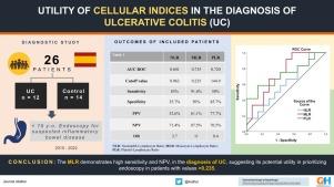
Diagnostic study of patients under 15 years of age undergoing colonoscopy ± esophagogastroduodenoscopy for suspected inflammatory bowel disease between 2015 and 2022 in a pediatric hospital. Patients with normal biopsy and anatomopathological diagnosis of UC were included. Using the area under the ROC curve, the values of platelet–lymphocyte ratio (PLR), neutrophil–lymphocyte ratio (NLR) and monocyte–lymphocyte ratio (MLR), calculated by dividing the number of platelets, neutrophils, and monocytes by the number of lymphocytes, respectively, were compared to establish the sensitivity, specificity, predictive values and odds ratio of each parameter in the diagnosis of UC.

Results

Twenty-six patients were included: 14 with normal biopsy and 12 with UC. PLR and MLR values were significantly higher in patients with UC (p < 0.05). The sensitivity, specificity, and negative predictive value of PLR, NLR and MLR for diagnosing UC were 58%, 83% and 91%; 85%, 35% and 50%; 70%, 71% and 87%, respectively. The biomarker with the highest diagnostic performance was MLR with a cutoff point of 0.235, area under the curve of 0.735 and odds ratio of 11 (95% CI 1.1–109.6; p = 0.041).

Conclusions

MLR has a high sensitivity, negative predictive value, and odds ratio in the pre-endoscopic diagnosis of ulcerative colitis. These findings, although exploratory, suggest that MLR could be useful in clinical practice in the initial diagnostic workup of UC, and perhaps in the future in the prioritization of endoscopic studies according to MLR values.

Abstract Image

探讨细胞指标在溃疡性结肠炎诊断中的应用
目的探讨细胞指标在溃疡性结肠炎(UC)诊断中的价值。方法对某儿科医院2015 - 2022年因疑似炎症性肠病行结肠镜检查±食管胃十二指肠镜检查的15岁以下患者进行诊断研究。包括活检和解剖病理诊断为UC的患者。利用ROC曲线下面积,将血小板-淋巴细胞比(PLR)、中性粒细胞-淋巴细胞比(NLR)、单核细胞-淋巴细胞比(MLR)分别以血小板、中性粒细胞、单核细胞数除以淋巴细胞数计算,比较各参数对UC诊断的敏感性、特异性、预测值和优势比。结果本组共纳入26例患者:14例活检正常,12例UC。UC患者的PLR和MLR值显著高于UC患者(p < 0.05)。PLR、NLR和MLR诊断UC的敏感性、特异性和阴性预测值分别为58%、83%和91%;85%, 35%和50%;分别为70%、71%和87%。诊断效能最高的生物标志物为MLR,截断点为0.235,曲线下面积为0.735,优势比为11 (95% CI 1.1 ~ 109.6; p = 0.041)。结论smlr对溃疡性结肠炎的内镜前诊断具有较高的敏感性、阴性预测值和优势比。这些发现虽然是探索性的,但表明在UC的初步诊断中,MLR可能在临床实践中有用,并且可能在未来根据MLR值确定内镜研究的优先级。
本文章由计算机程序翻译,如有差异,请以英文原文为准。
求助全文
约1分钟内获得全文 求助全文
来源期刊
自引率
0.00%
发文量
0
×
引用
GB/T 7714-2015
复制
MLA
复制
APA
复制
导出至
BibTeX EndNote RefMan NoteFirst NoteExpress
×
提示
您的信息不完整,为了账户安全,请先补充。
现在去补充
×
提示
您因"违规操作"
具体请查看互助需知
我知道了
×
提示
确定
请完成安全验证×
copy
已复制链接
快去分享给好友吧!
我知道了
右上角分享
点击右上角分享
0
联系我们:info@booksci.cn Book学术提供免费学术资源搜索服务,方便国内外学者检索中英文文献。致力于提供最便捷和优质的服务体验。 Copyright © 2023 布克学术 All rights reserved.
京ICP备2023020795号-1
ghs 京公网安备 11010802042870号
Book学术文献互助
Book学术文献互助群
群 号:604180095
Book学术官方微信