Tatielly Karine Costa Alves, Bruna Lara França Lima, Ana Clara Gonzaga da Costa Ferreira, Giulio Cesar Moreira Manzi, Franca Arenare Jeunon, Micena Roberta Miranda Alves E Silva, Flávio Ricardo Manzi
{"title":"Influence of cone-beam computed tomography reconstruction parameters on bone fractal dimension: A cross-sectional observational study.","authors":"Tatielly Karine Costa Alves, Bruna Lara França Lima, Ana Clara Gonzaga da Costa Ferreira, Giulio Cesar Moreira Manzi, Franca Arenare Jeunon, Micena Roberta Miranda Alves E Silva, Flávio Ricardo Manzi","doi":"10.5624/isd.20250018","DOIUrl":null,"url":null,"abstract":"<p><strong>Purpose: </strong>This study aimed to evaluate the influence of different cone-beam computed tomography (CBCT) image reconstruction parameters (slice thickness, noise filter application and orthogonal plane) on the calculation of bone fractal dimension and, based on those findings, to determine the optimal protocol for this type of assessment.</p><p><strong>Materials and methods: </strong>The sample consisted of 18 patients who underwent CBCT scans of the mandible and bone densitometry examinations. Four mandibular regions of interest were selected from the scans, with various image reconstruction parameters applied. Fractal dimension was calculated using the box-counting method. Two independent observers performed the evaluations, and all analyses were conducted with a significance level of 5%.</p><p><strong>Results: </strong>The retromolar triangle and mandibular body regions did not demonstrate statistically significant differences when different tomographic reconstruction parameters were applied (<i>P</i>>0.05). The mandibular base did not display a consistent pattern that could define the influence of these parameters on its evaluation. The symphysis region showed improved performance in fractal analysis when using sagittal plane images with a 1 mm slice thickness.</p><p><strong>Conclusion: </strong>Operator-dependent parameters inherent to navigation software can influence fractal dimension analysis, with variations depending on the region of interest. The most appropriate parameters for this evaluation were identified as the sagittal plane with a 1 mm slice thickness. Among the regions assessed, the mandibular body was found to be the most suitable for fractal dimension analysis in CBCT.</p>","PeriodicalId":51714,"journal":{"name":"Imaging Science in Dentistry","volume":"55 3","pages":"261-270"},"PeriodicalIF":2.1000,"publicationDate":"2025-09-01","publicationTypes":"Journal Article","fieldsOfStudy":null,"isOpenAccess":false,"openAccessPdf":"https://www.ncbi.nlm.nih.gov/pmc/articles/PMC12505442/pdf/","citationCount":"0","resultStr":null,"platform":"Semanticscholar","paperid":null,"PeriodicalName":"Imaging Science in Dentistry","FirstCategoryId":"1085","ListUrlMain":"https://doi.org/10.5624/isd.20250018","RegionNum":0,"RegionCategory":null,"ArticlePicture":[],"TitleCN":null,"AbstractTextCN":null,"PMCID":null,"EPubDate":"2025/7/12 0:00:00","PubModel":"Epub","JCR":"Q3","JCRName":"DENTISTRY, ORAL SURGERY & MEDICINE","Score":null,"Total":0}
引用次数: 0
Abstract
Purpose: This study aimed to evaluate the influence of different cone-beam computed tomography (CBCT) image reconstruction parameters (slice thickness, noise filter application and orthogonal plane) on the calculation of bone fractal dimension and, based on those findings, to determine the optimal protocol for this type of assessment.
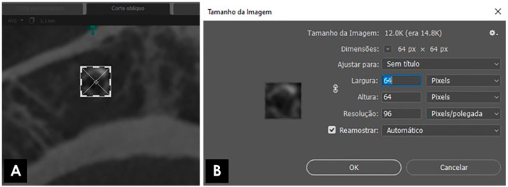
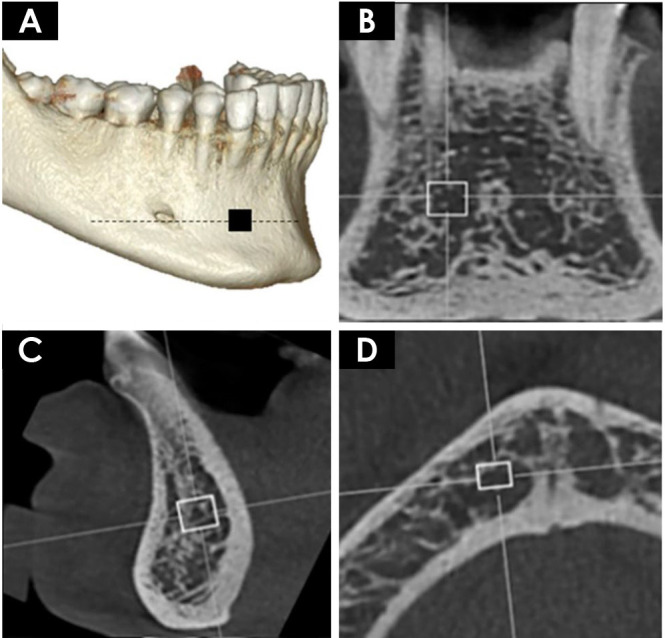
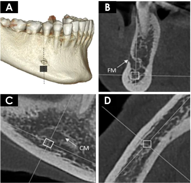
Materials and methods: The sample consisted of 18 patients who underwent CBCT scans of the mandible and bone densitometry examinations. Four mandibular regions of interest were selected from the scans, with various image reconstruction parameters applied. Fractal dimension was calculated using the box-counting method. Two independent observers performed the evaluations, and all analyses were conducted with a significance level of 5%.
Results: The retromolar triangle and mandibular body regions did not demonstrate statistically significant differences when different tomographic reconstruction parameters were applied (P>0.05). The mandibular base did not display a consistent pattern that could define the influence of these parameters on its evaluation. The symphysis region showed improved performance in fractal analysis when using sagittal plane images with a 1 mm slice thickness.
Conclusion: Operator-dependent parameters inherent to navigation software can influence fractal dimension analysis, with variations depending on the region of interest. The most appropriate parameters for this evaluation were identified as the sagittal plane with a 1 mm slice thickness. Among the regions assessed, the mandibular body was found to be the most suitable for fractal dimension analysis in CBCT.

 求助内容:
求助内容: 应助结果提醒方式:


