Cone-beam computed tomography-based analysis of jawbone destruction patterns in multiple myeloma: Associations with clinical data in an observational study.
Thaiza Goncalves Rocha, Raphael Dos Santos Alves Martins Veiga, Eduardo Murad Villoria, Roberto Josè Pessoa de Magalhães Filho, Angelo Maiolino, Sandra Regina Torres, Maria Augusta Visconti
{"title":"Cone-beam computed tomography-based analysis of jawbone destruction patterns in multiple myeloma: Associations with clinical data in an observational study.","authors":"Thaiza Goncalves Rocha, Raphael Dos Santos Alves Martins Veiga, Eduardo Murad Villoria, Roberto Josè Pessoa de Magalhães Filho, Angelo Maiolino, Sandra Regina Torres, Maria Augusta Visconti","doi":"10.5624/isd.20250015","DOIUrl":null,"url":null,"abstract":"<p><strong>Purpose: </strong>This study analyzed cone-beam computed tomography images of 27 patients with multiple myeloma at different disease stages to identify jawbone destruction patterns and assess their associations with clinical data.</p><p><strong>Materials and methods: </strong>In this cross-sectional study, 2 trained examiners performed standardized, consensus-based image analyses. Lesions were classified into 4 distinct bone destruction patterns: diffuse, multilocular, unilocular, and punched-out. Clinical data were collected from medical records.</p><p><strong>Results: </strong>The sample included 51.8% male and 48.2% female patients, predominantly between 42 and 60 years old. All cases exhibited diffuse bone destruction affecting both jaws. Multilocular and unilocular patterns were observed in 51.9% and 29.6% of cases, respectively, while no punched-out lesions were identified. The unilocular pattern was significantly associated with cases classified as International Staging System stage I and Durie-Salmon stage IIIA.</p><p><strong>Conclusion: </strong>Among the studied cases of multiple myeloma, the most frequently observed bone destruction patterns were diffuse and multilocular. The absence of punched-out lesions may be attributable to the use of 3-dimensional imaging. A clear association was identified between the unilocular pattern and disease staging.</p>","PeriodicalId":51714,"journal":{"name":"Imaging Science in Dentistry","volume":"55 3","pages":"253-260"},"PeriodicalIF":2.1000,"publicationDate":"2025-09-01","publicationTypes":"Journal Article","fieldsOfStudy":null,"isOpenAccess":false,"openAccessPdf":"https://www.ncbi.nlm.nih.gov/pmc/articles/PMC12505440/pdf/","citationCount":"0","resultStr":null,"platform":"Semanticscholar","paperid":null,"PeriodicalName":"Imaging Science in Dentistry","FirstCategoryId":"1085","ListUrlMain":"https://doi.org/10.5624/isd.20250015","RegionNum":0,"RegionCategory":null,"ArticlePicture":[],"TitleCN":null,"AbstractTextCN":null,"PMCID":null,"EPubDate":"2025/7/1 0:00:00","PubModel":"Epub","JCR":"Q3","JCRName":"DENTISTRY, ORAL SURGERY & MEDICINE","Score":null,"Total":0}
引用次数: 0
Abstract
Purpose: This study analyzed cone-beam computed tomography images of 27 patients with multiple myeloma at different disease stages to identify jawbone destruction patterns and assess their associations with clinical data.
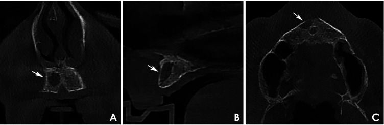
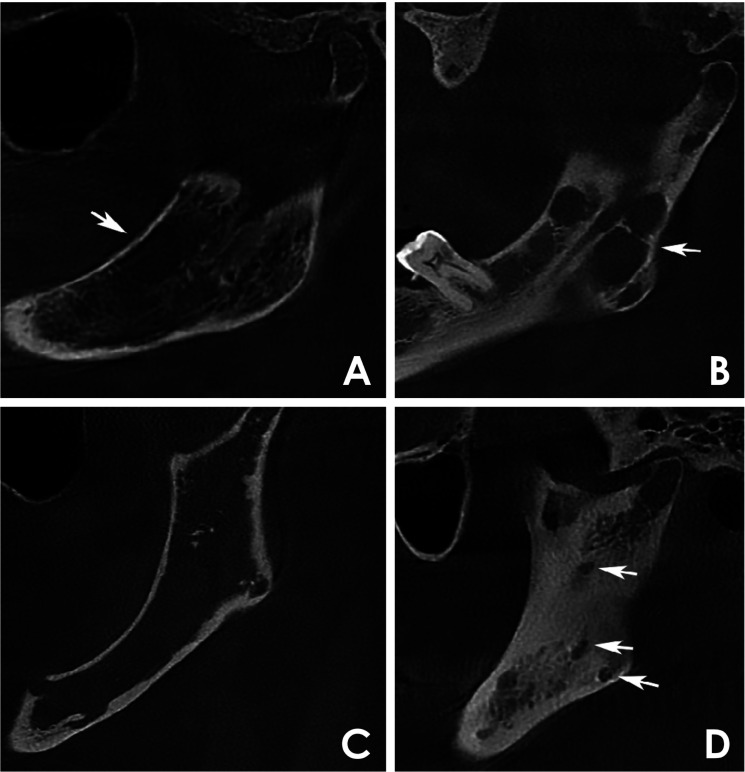
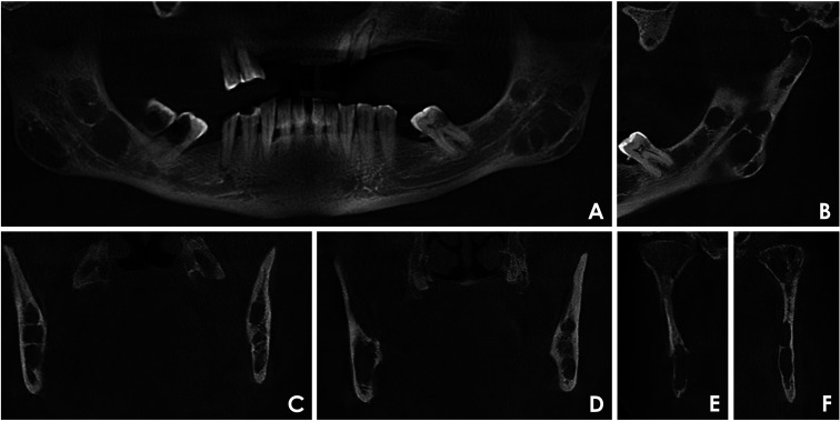
Materials and methods: In this cross-sectional study, 2 trained examiners performed standardized, consensus-based image analyses. Lesions were classified into 4 distinct bone destruction patterns: diffuse, multilocular, unilocular, and punched-out. Clinical data were collected from medical records.
Results: The sample included 51.8% male and 48.2% female patients, predominantly between 42 and 60 years old. All cases exhibited diffuse bone destruction affecting both jaws. Multilocular and unilocular patterns were observed in 51.9% and 29.6% of cases, respectively, while no punched-out lesions were identified. The unilocular pattern was significantly associated with cases classified as International Staging System stage I and Durie-Salmon stage IIIA.
Conclusion: Among the studied cases of multiple myeloma, the most frequently observed bone destruction patterns were diffuse and multilocular. The absence of punched-out lesions may be attributable to the use of 3-dimensional imaging. A clear association was identified between the unilocular pattern and disease staging.

 求助内容:
求助内容: 应助结果提醒方式:


