Marco Serafin, Benedetta Baldini, Elisa Boccalari, Francesca Parravicini, Piero Antonio Zecca, Alberto Caprioglio
{"title":"Accuracy of facial scan registration: A comparison between full-cranium and reduced field-of-view cone-beam computed tomography.","authors":"Marco Serafin, Benedetta Baldini, Elisa Boccalari, Francesca Parravicini, Piero Antonio Zecca, Alberto Caprioglio","doi":"10.5624/isd.20250013","DOIUrl":null,"url":null,"abstract":"<p><strong>Purpose: </strong>This retrospective study aimed to evaluate the accuracy of facial scan (FS) to cone-beam computed tomography (CBCT) registration by comparing superimpositions on full-cranium and reduced field-of-view (FOV) CBCT, with the goal of assessing its potential to reduce radiation exposure without compromising diagnostic quality.</p><p><strong>Materials and methods: </strong>CBCT scans from 50 patients were analyzed, integrating FS data obtained via 3D laser scanning. FSs were registered to both full-cranium and reduced FOV CBCT using landmark-based matching and a best-fit algorithm. Accuracy was evaluated by calculating the point-to-point surface distance between FS and CBCT soft-tissue renderings. The metrics used were root mean square distance (RMSD), Hausdorff distance (HD), and median distance (MD). Registration of FS onto full FOV CBCT served as the ground truth. Statistical analysis employed the Mann-Whitney U test to compare registration performance on both the overall surface and the facial midline.</p><p><strong>Results: </strong>There was no significant difference in HD (<i>P</i>=0.288) between the 2 methods. However, median RMSD and MD were significantly lower for full-cranium CBCT (<i>P</i>=0.019). Midline alignment between FS and reduced FOV CBCT showed no visual discrepancies, with an MD of 0.35 mm along the midsagittal plane.</p><p><strong>Conclusion: </strong>FS registration to reduced FOV CBCT provides clinically acceptable accuracy, particularly in the midline region, while substantially reducing radiation exposure. This approach is promising for a range of dental applications, especially in pediatric cases and situations prioritizing facial aesthetics. Further research is warranted to optimize this technique for diverse clinical contexts.</p>","PeriodicalId":51714,"journal":{"name":"Imaging Science in Dentistry","volume":"55 3","pages":"245-252"},"PeriodicalIF":2.1000,"publicationDate":"2025-09-01","publicationTypes":"Journal Article","fieldsOfStudy":null,"isOpenAccess":false,"openAccessPdf":"https://www.ncbi.nlm.nih.gov/pmc/articles/PMC12505441/pdf/","citationCount":"0","resultStr":null,"platform":"Semanticscholar","paperid":null,"PeriodicalName":"Imaging Science in Dentistry","FirstCategoryId":"1085","ListUrlMain":"https://doi.org/10.5624/isd.20250013","RegionNum":0,"RegionCategory":null,"ArticlePicture":[],"TitleCN":null,"AbstractTextCN":null,"PMCID":null,"EPubDate":"2025/7/12 0:00:00","PubModel":"Epub","JCR":"Q3","JCRName":"DENTISTRY, ORAL SURGERY & MEDICINE","Score":null,"Total":0}
引用次数: 0
Abstract
Purpose: This retrospective study aimed to evaluate the accuracy of facial scan (FS) to cone-beam computed tomography (CBCT) registration by comparing superimpositions on full-cranium and reduced field-of-view (FOV) CBCT, with the goal of assessing its potential to reduce radiation exposure without compromising diagnostic quality.
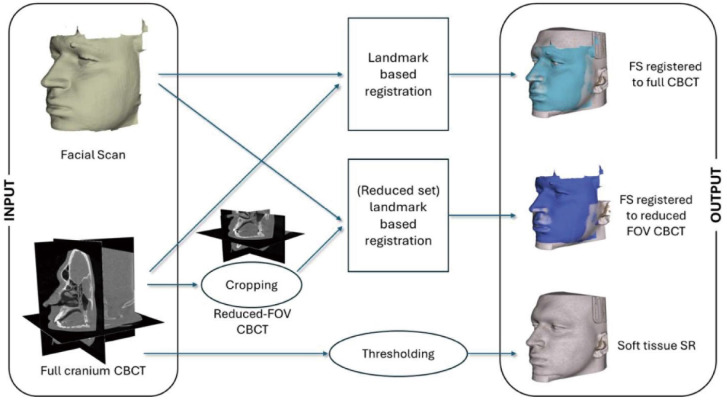
Materials and methods: CBCT scans from 50 patients were analyzed, integrating FS data obtained via 3D laser scanning. FSs were registered to both full-cranium and reduced FOV CBCT using landmark-based matching and a best-fit algorithm. Accuracy was evaluated by calculating the point-to-point surface distance between FS and CBCT soft-tissue renderings. The metrics used were root mean square distance (RMSD), Hausdorff distance (HD), and median distance (MD). Registration of FS onto full FOV CBCT served as the ground truth. Statistical analysis employed the Mann-Whitney U test to compare registration performance on both the overall surface and the facial midline.
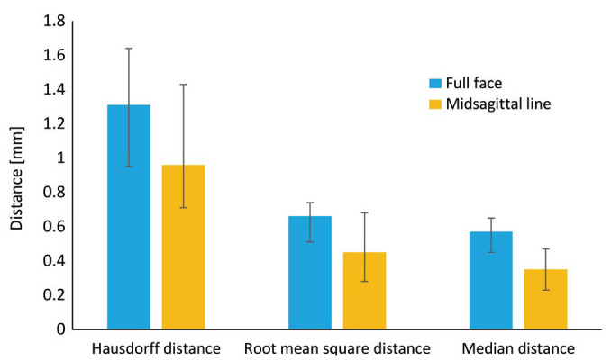
Results: There was no significant difference in HD (P=0.288) between the 2 methods. However, median RMSD and MD were significantly lower for full-cranium CBCT (P=0.019). Midline alignment between FS and reduced FOV CBCT showed no visual discrepancies, with an MD of 0.35 mm along the midsagittal plane.
Conclusion: FS registration to reduced FOV CBCT provides clinically acceptable accuracy, particularly in the midline region, while substantially reducing radiation exposure. This approach is promising for a range of dental applications, especially in pediatric cases and situations prioritizing facial aesthetics. Further research is warranted to optimize this technique for diverse clinical contexts.

 求助内容:
求助内容: 应助结果提醒方式:


