{"title":"Osteogenic effects of metformin and exenatide on bone regeneration in non-diabetic rats: A Micro-CT and histological study.","authors":"Hasan Ozturk, Neslihan Simsek, Levent Akinci, Meltem Ozgocmen, Dilek Helvacioglu Yigit","doi":"10.4103/jomfp.jomfp_4_25","DOIUrl":null,"url":null,"abstract":"<p><strong>Aim: </strong>This study aimed to evaluate the osteogenic effects of systemic metformin and exenatide administration on bone tissue regeneration in an experimental rat model by utilising micro-computed tomography (micro-CT) and histological analysis.</p><p><strong>Materials and methods: </strong>Uniform craniotomy defects measuring 3 mm in diameter and 2 mm depth were performed in the parietal bones of 27 female albino Wistar rats, which were randomly divided into three groups: 1) a group receiving 100 mg/kg/day of oral metformin, 2) a group receiving 3 μg/kg/day of intraperitoneal exenatide, and 3) a control group receiving no medication. Bone volume and density at the defect site were evaluated using micro-CT scanning and analysis.</p><p><strong>Results: </strong>Bone regeneration and the integration of newly formed bone into intact bone were assessed through histological and immunohistochemical examinations. In all three groups, the results showed no significant differences in bone volume, bone density, the presence of fibrous connective tissue, or the complete integration of the defect area into the bone tissue. However, the experimental groups exhibited significant differences in the number of osteoblasts (<i>P</i> = 0.007) and osteoclasts (<i>P</i> = 0.007) when compared to the control group.</p><p><strong>Conclusions: </strong>Metformin and exenatide enhance the activity of osteoblasts and osteoclasts in bone defects, promoting osteogenic potential during the healing process in non-diabetic rats.</p>","PeriodicalId":38846,"journal":{"name":"Journal of Oral and Maxillofacial Pathology","volume":"29 3","pages":"352-359"},"PeriodicalIF":0.0000,"publicationDate":"2025-07-01","publicationTypes":"Journal Article","fieldsOfStudy":null,"isOpenAccess":false,"openAccessPdf":"https://www.ncbi.nlm.nih.gov/pmc/articles/PMC12507372/pdf/","citationCount":"0","resultStr":null,"platform":"Semanticscholar","paperid":null,"PeriodicalName":"Journal of Oral and Maxillofacial Pathology","FirstCategoryId":"1085","ListUrlMain":"https://doi.org/10.4103/jomfp.jomfp_4_25","RegionNum":0,"RegionCategory":null,"ArticlePicture":[],"TitleCN":null,"AbstractTextCN":null,"PMCID":null,"EPubDate":"2025/9/26 0:00:00","PubModel":"Epub","JCR":"Q3","JCRName":"Medicine","Score":null,"Total":0}
引用次数: 0
Abstract
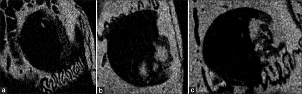
Aim: This study aimed to evaluate the osteogenic effects of systemic metformin and exenatide administration on bone tissue regeneration in an experimental rat model by utilising micro-computed tomography (micro-CT) and histological analysis.
Materials and methods: Uniform craniotomy defects measuring 3 mm in diameter and 2 mm depth were performed in the parietal bones of 27 female albino Wistar rats, which were randomly divided into three groups: 1) a group receiving 100 mg/kg/day of oral metformin, 2) a group receiving 3 μg/kg/day of intraperitoneal exenatide, and 3) a control group receiving no medication. Bone volume and density at the defect site were evaluated using micro-CT scanning and analysis.
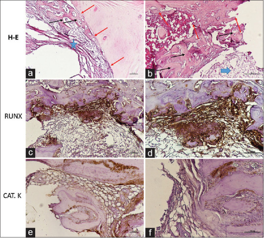
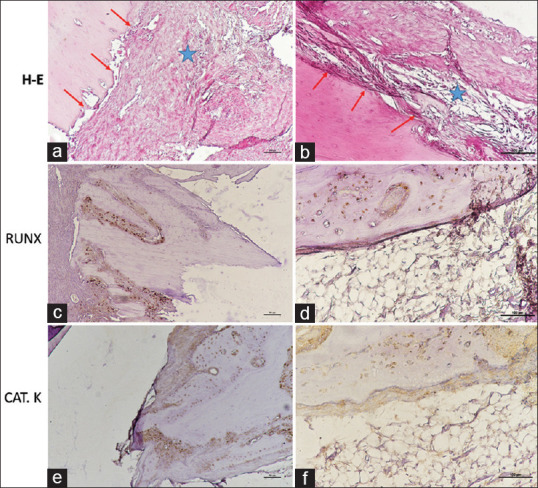
Results: Bone regeneration and the integration of newly formed bone into intact bone were assessed through histological and immunohistochemical examinations. In all three groups, the results showed no significant differences in bone volume, bone density, the presence of fibrous connective tissue, or the complete integration of the defect area into the bone tissue. However, the experimental groups exhibited significant differences in the number of osteoblasts (P = 0.007) and osteoclasts (P = 0.007) when compared to the control group.
Conclusions: Metformin and exenatide enhance the activity of osteoblasts and osteoclasts in bone defects, promoting osteogenic potential during the healing process in non-diabetic rats.
期刊介绍:
The journal of Oral and Maxillofacial Pathology [ISSN:print-(0973-029X, online-1998-393X)] is a tri-annual journal published on behalf of “The Indian Association of Oral and Maxillofacial Pathologists” (IAOMP). The publication of JOMFP was started in the year 1993. The journal publishes papers on a wide spectrum of topics associated with the scope of Oral and Maxillofacial Pathology, also, ensuring scientific merit and quality. It is a comprehensive reading material for the professionals who want to upgrade their diagnostic skills in Oral Diseases; allows exposure to newer topics and methods of research in the Oral-facial Tissues and Pathology. New features allow an open minded thinking and approach to various pathologies. It also encourages authors to showcase quality work done by them and to compile relevant cases which are diagnostically challenging. The Journal takes pride in maintaining the quality of articles and photomicrographs.

 求助内容:
求助内容: 应助结果提醒方式:


