Khadijah Mohideen, Chadrasekaran Krithika, Nadeem Jeddy, Pratibha Ramani, Mutaz Ali Hasan, Uma Sudhakar
{"title":"Evaluation of the immunohistochemical expression of oxidative DNA damage marker 8-OHdG in oral cancer.","authors":"Khadijah Mohideen, Chadrasekaran Krithika, Nadeem Jeddy, Pratibha Ramani, Mutaz Ali Hasan, Uma Sudhakar","doi":"10.4103/jomfp.jomfp_169_25","DOIUrl":null,"url":null,"abstract":"<p><strong>Objective: </strong>The present immunohistochemical investigation attempted to evaluate the concentrations of 8-hydroxy-2-deoxyguanosine (8-OHdG) as a prospective DNA damage biomarker in primary oral squamous cell carcinoma (OSCC) patients compared to healthy tissues to analyse the immunohistochemistry (IHC) staining intensity levels across different histopathological levels of OSCC.</p><p><strong>Materials and methods: </strong>The control group consisted of tissue samples from the normal mucosa (n = 10). The study groups consisted of tissue blocks sourced from patients with primary OSCC (n = 55). The principal antibody utilised was 8-OHdG Antibody, and a control specimen was treated identically to the OSCC group but treated in non-immune serum rather than the primary antibody. The data were estimated using the Statistical Package for the Social Sciences (SPSS) version 26.0. The Fisher's exact test was employed to examine the variations between the sample groups at a 5% significance level.</p><p><strong>Results: </strong>The OSCC group had 20 well-differentiated (WD), 20 moderately differentiated (MD), and 15 poorly differentiated (PD) samples. Compared to normal tissue samples, a marked elevation of 8-OHdG antigen production was observed in OSCC tissue samples (<i>P</i> < 0.0001). The WD cohort showed moderate expression of the 8-OHdG antigen in 50%, mild in 30%, and intense in 20%. In MD samples, 5% showed intense expression, moderate in 55%, and mild staining in 40%. In PD samples, 46.67% showed mild and moderate 8-OHdG expression, whereas 6.66% showed intense expression. However, the difference was insignificant (<i>P</i> > 0.05).</p><p><strong>Conclusion: </strong>The Oxidative stress (OS) marker 8-OHdG has higher concentrations in the tissue samples with OSCC than in healthy tissues and serves as a predictive biomarker for OSCC.</p>","PeriodicalId":38846,"journal":{"name":"Journal of Oral and Maxillofacial Pathology","volume":"29 3","pages":"458-464"},"PeriodicalIF":0.0000,"publicationDate":"2025-07-01","publicationTypes":"Journal Article","fieldsOfStudy":null,"isOpenAccess":false,"openAccessPdf":"https://www.ncbi.nlm.nih.gov/pmc/articles/PMC12507358/pdf/","citationCount":"0","resultStr":null,"platform":"Semanticscholar","paperid":null,"PeriodicalName":"Journal of Oral and Maxillofacial Pathology","FirstCategoryId":"1085","ListUrlMain":"https://doi.org/10.4103/jomfp.jomfp_169_25","RegionNum":0,"RegionCategory":null,"ArticlePicture":[],"TitleCN":null,"AbstractTextCN":null,"PMCID":null,"EPubDate":"2025/9/26 0:00:00","PubModel":"Epub","JCR":"Q3","JCRName":"Medicine","Score":null,"Total":0}
引用次数: 0
Abstract
Objective: The present immunohistochemical investigation attempted to evaluate the concentrations of 8-hydroxy-2-deoxyguanosine (8-OHdG) as a prospective DNA damage biomarker in primary oral squamous cell carcinoma (OSCC) patients compared to healthy tissues to analyse the immunohistochemistry (IHC) staining intensity levels across different histopathological levels of OSCC.
Materials and methods: The control group consisted of tissue samples from the normal mucosa (n = 10). The study groups consisted of tissue blocks sourced from patients with primary OSCC (n = 55). The principal antibody utilised was 8-OHdG Antibody, and a control specimen was treated identically to the OSCC group but treated in non-immune serum rather than the primary antibody. The data were estimated using the Statistical Package for the Social Sciences (SPSS) version 26.0. The Fisher's exact test was employed to examine the variations between the sample groups at a 5% significance level.
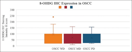
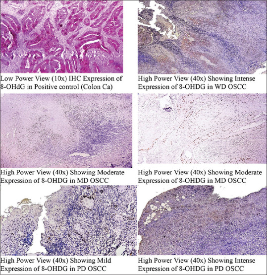
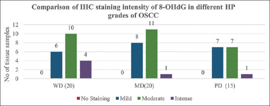
Results: The OSCC group had 20 well-differentiated (WD), 20 moderately differentiated (MD), and 15 poorly differentiated (PD) samples. Compared to normal tissue samples, a marked elevation of 8-OHdG antigen production was observed in OSCC tissue samples (P < 0.0001). The WD cohort showed moderate expression of the 8-OHdG antigen in 50%, mild in 30%, and intense in 20%. In MD samples, 5% showed intense expression, moderate in 55%, and mild staining in 40%. In PD samples, 46.67% showed mild and moderate 8-OHdG expression, whereas 6.66% showed intense expression. However, the difference was insignificant (P > 0.05).
Conclusion: The Oxidative stress (OS) marker 8-OHdG has higher concentrations in the tissue samples with OSCC than in healthy tissues and serves as a predictive biomarker for OSCC.
期刊介绍:
The journal of Oral and Maxillofacial Pathology [ISSN:print-(0973-029X, online-1998-393X)] is a tri-annual journal published on behalf of “The Indian Association of Oral and Maxillofacial Pathologists” (IAOMP). The publication of JOMFP was started in the year 1993. The journal publishes papers on a wide spectrum of topics associated with the scope of Oral and Maxillofacial Pathology, also, ensuring scientific merit and quality. It is a comprehensive reading material for the professionals who want to upgrade their diagnostic skills in Oral Diseases; allows exposure to newer topics and methods of research in the Oral-facial Tissues and Pathology. New features allow an open minded thinking and approach to various pathologies. It also encourages authors to showcase quality work done by them and to compile relevant cases which are diagnostically challenging. The Journal takes pride in maintaining the quality of articles and photomicrographs.

 求助内容:
求助内容: 应助结果提醒方式:


