Tommaso Calzoni, Graziella Donatelli, Gianmichele Migaleddu, Marta Lancione, Paolo Cecchi, Laura Biagi, Michele Caniglia, Roberto Ceravolo, Mirco Cosottini
{"title":"Hypertrophic olivary degeneration: a 7 Tesla advanced imaging case report.","authors":"Tommaso Calzoni, Graziella Donatelli, Gianmichele Migaleddu, Marta Lancione, Paolo Cecchi, Laura Biagi, Michele Caniglia, Roberto Ceravolo, Mirco Cosottini","doi":"10.3389/fnins.2025.1656655","DOIUrl":null,"url":null,"abstract":"<p><strong>Background and objectives: </strong>A 50-year-old patient developed ataxia, nystagmus, and palatal tremor. Conventional magnetic resonance imaging (MRI) revealed inferior olivary nuclei enlargement and hyperintensity in T2-weighted images, indicating hypertrophic olivary degeneration (HOD). The patient's past medical history reported proton therapy for an VIII cranial nerve Schwannoma. Here, we aimed to investigate the potential alterations involving tracts and nuclei composing the dentato-rubro-olivary pathway (Guillain-Mollaret triangle) using an advanced ultra-high field (7 T) MRI protocol.</p><p><strong>Materials and methods: </strong>The patient underwent a 7 T-MRI brain exam, including a multi-echo gradient-echo sequence for quantitative susceptibility mapping and diffusion tensor imaging (DTI). The DTI dataset was elaborated for tractography and computation of tensor metrics.</p><p><strong>Results: </strong>7 T-MRI allowed the depiction of the brainstem tracts and nuclei composing the Guillain-Mollaret triangle. Both qualitative and quantitative analyses of these structures demonstrated damage to the right red nucleus and the dentato-rubral tracts bilaterally. These findings are consistent with the pathophysiology of HOD and were confirmed in a follow-up MRI.</p><p><strong>Discussion: </strong>This study highlights the capability of 7 T-MRI to depict and investigate brainstem substructures such as tracts and nuclei. To the best of our knowledge, this is the first study to depict all tracts composing the Guillain-Mollaret triangle and directly document their alterations in HOD.</p>","PeriodicalId":12639,"journal":{"name":"Frontiers in Neuroscience","volume":"19 ","pages":"1656655"},"PeriodicalIF":3.2000,"publicationDate":"2025-09-24","publicationTypes":"Journal Article","fieldsOfStudy":null,"isOpenAccess":false,"openAccessPdf":"https://www.ncbi.nlm.nih.gov/pmc/articles/PMC12504213/pdf/","citationCount":"0","resultStr":null,"platform":"Semanticscholar","paperid":null,"PeriodicalName":"Frontiers in Neuroscience","FirstCategoryId":"3","ListUrlMain":"https://doi.org/10.3389/fnins.2025.1656655","RegionNum":3,"RegionCategory":"医学","ArticlePicture":[],"TitleCN":null,"AbstractTextCN":null,"PMCID":null,"EPubDate":"2025/1/1 0:00:00","PubModel":"eCollection","JCR":"Q2","JCRName":"NEUROSCIENCES","Score":null,"Total":0}
引用次数: 0
Abstract
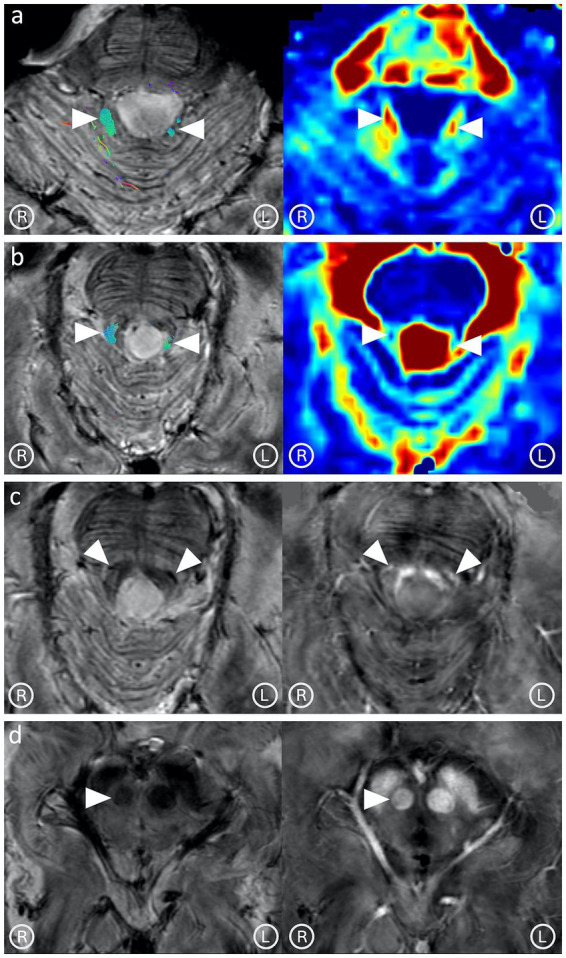
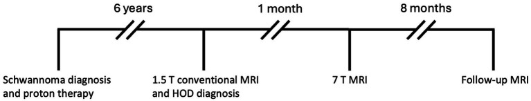
Background and objectives: A 50-year-old patient developed ataxia, nystagmus, and palatal tremor. Conventional magnetic resonance imaging (MRI) revealed inferior olivary nuclei enlargement and hyperintensity in T2-weighted images, indicating hypertrophic olivary degeneration (HOD). The patient's past medical history reported proton therapy for an VIII cranial nerve Schwannoma. Here, we aimed to investigate the potential alterations involving tracts and nuclei composing the dentato-rubro-olivary pathway (Guillain-Mollaret triangle) using an advanced ultra-high field (7 T) MRI protocol.
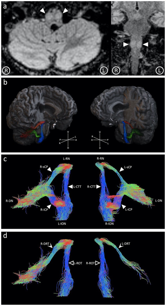
Materials and methods: The patient underwent a 7 T-MRI brain exam, including a multi-echo gradient-echo sequence for quantitative susceptibility mapping and diffusion tensor imaging (DTI). The DTI dataset was elaborated for tractography and computation of tensor metrics.
Results: 7 T-MRI allowed the depiction of the brainstem tracts and nuclei composing the Guillain-Mollaret triangle. Both qualitative and quantitative analyses of these structures demonstrated damage to the right red nucleus and the dentato-rubral tracts bilaterally. These findings are consistent with the pathophysiology of HOD and were confirmed in a follow-up MRI.
Discussion: This study highlights the capability of 7 T-MRI to depict and investigate brainstem substructures such as tracts and nuclei. To the best of our knowledge, this is the first study to depict all tracts composing the Guillain-Mollaret triangle and directly document their alterations in HOD.
期刊介绍:
Neural Technology is devoted to the convergence between neurobiology and quantum-, nano- and micro-sciences. In our vision, this interdisciplinary approach should go beyond the technological development of sophisticated methods and should contribute in generating a genuine change in our discipline.
 求助内容:
求助内容: 应助结果提醒方式:


