Regional and cell type-specific activation of the unfolded protein response after kainate injection in mice
IF 4
3区 医学
Q2 BIOCHEMISTRY & MOLECULAR BIOLOGY
引用次数: 0
Abstract
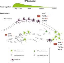
The unfolded protein response (UPR) is activated under different neuropathological conditions, such as brain ischemia, epilepsy, and neurodegeneration. We previously reported that a UPR transducer, activating transcription factor 6 (ATF6), and its downstream molecular chaperones in the endoplasmic reticulum (ER) have neuroprotective properties against excitotoxicity. In this study, we examined the temporal and spatial changes in the UPR activation after administration of an excitotoxic reagent, kainate (KA), into mice. RT-qPCR revealed enhanced expression of UPR genes, with peaks either on day 1 or day 3 after intrahippocampal KA injection. The status of the UPR was analyzed using ER stress-activated indicator (ERAI)-transgenic mice, in which the spliced form of XBP-1, downstream of the IRE1 branch of the UPR, can be monitored. ERAI-derived GFP signals were strongly observed in CA3 neurons and moderately observed in dentate gyrus neurons, but not in CA1 neurons, after KA injection. A small portion of the activated astrocytes was also positive for ERAI signals. Further studies revealed that ERAI signals were observed in both the soma and dendrites of neurons in regions with enhanced neuronal activity and resistance to KA toxicity. These results suggest that the UPR may be associated with the neuronal activity and survival after KA injection.
海碱盐注射后未折叠蛋白反应的区域和细胞特异性激活。
未折叠蛋白反应(UPR)在不同的神经病理条件下被激活,如脑缺血、癫痫和神经变性。我们之前报道了UPR传感器,激活转录因子6 (ATF6)及其下游内质网(ER)中的分子伴侣具有抗兴奋性毒性的神经保护特性。在这项研究中,我们检测了给药兴奋毒性试剂kainate (KA)后小鼠UPR激活的时间和空间变化。RT-qPCR显示UPR基因表达增强,在海马内注射KA后第1天或第3天达到峰值。利用ER应激激活指示剂(ERAI)转基因小鼠分析了UPR的状态,其中可以监测UPR IRE1分支下游的XBP-1的剪接形式。KA注射后,在CA3神经元中观察到erai衍生的GFP信号强烈,在齿状回神经元中观察到中度,但在CA1神经元中没有。小部分活化的星形胶质细胞ERAI信号也呈阳性。进一步的研究表明,ERAI信号在神经元活性增强和KA毒性抵抗区域的神经元体细胞和树突中都观察到。这些结果提示,KA注射后的UPR可能与神经元活动和存活有关。
本文章由计算机程序翻译,如有差异,请以英文原文为准。
求助全文
约1分钟内获得全文
求助全文
来源期刊
Neurochemistry international
医学-神经科学
CiteScore
8.40
自引率
2.40%
发文量
128
审稿时长
37 days
期刊介绍:
Neurochemistry International is devoted to the rapid publication of outstanding original articles and timely reviews in neurochemistry. Manuscripts on a broad range of topics will be considered, including molecular and cellular neurochemistry, neuropharmacology and genetic aspects of CNS function, neuroimmunology, metabolism as well as the neurochemistry of neurological and psychiatric disorders of the CNS.
 求助内容:
求助内容: 应助结果提醒方式:


