Xuerou Li, Fuwen Dong, Xiaofei Chen, Xingxin Luo, Wenqi Wang
{"title":"Radiological frontiers in understanding paraspinal muscle pathophysiology in chronic low back pain.","authors":"Xuerou Li, Fuwen Dong, Xiaofei Chen, Xingxin Luo, Wenqi Wang","doi":"10.3389/fmed.2025.1653876","DOIUrl":null,"url":null,"abstract":"<p><strong>Background: </strong>Paraspinal muscles have a profound role in maintaining spinal stability and are often implicated in spinal degenerative conditions as well as chronic low back pain (CLBP). Alterations in these muscles have significant clinical implications for early prevention, treatment strategies, prognosis, and understanding the underlying mechanisms of CLBP. Recent advances in imaging techniques can generate prominent structural and functional characteristics of these muscles.</p><p><strong>Objectives: </strong>This study is specifically to review recent advancements in imaging techniques focusing on the regenerative and degenerative properties pertinent to paraspinal muscles in the context of CLBP.</p><p><strong>Methods: </strong>A literature review was executed to ascertain the databases including PubMed, Google Scholar, RelMed, and the National Library of Medicine. The search included studies elucidating recent imaging advancements, fiber-type composition analysis, level/depth-specific muscle characteristics, and clinical applications of novel radiological techniques in evaluating paraspinal muscle morphology and function. We performed this review without comprehensive meta-analysis.</p><p><strong>Results: </strong>The review identified significant advancements in imaging modalities for assessing paraspinal muscles, including functional MRI (fMRI), quantitative MRI (qMRI), and T2 mapping techniques. Key findings include: Fiber-type composition analysis: Recent studies elucidate the role of depth-dependent fiber-type gradients along with their correlation with muscle function in health and disease. Standardized imaging protocols: The lack of uniform imaging protocols remains a challenge, emphasizing the need for standardization to improve reproducibility and reliability. Radiological advances: Emerging techniques such as advanced fMRI and qMRI enable detailed visualization of muscle structure and function, overcoming limitations of traditional imaging methods. Age-related microvascular changes: age-related microvascular alterations significantly impact paraspinal muscle morphology and can be effectively captured by modern imaging biomarkers.</p><p><strong>Conclusion: </strong>Advances in imaging techniques have enhanced our understanding of the structural and functional changes in paraspinal muscles associated with CLBP. The integration of imaging biomarkers into clinical practice holds promise for early diagnosis, targeted interventions, and better prognostic evaluations. Future research should focus on developing standardized imaging protocols and further exploring depth-specific properties of paraspinal muscles to enhance clinical outcomes.</p>","PeriodicalId":12488,"journal":{"name":"Frontiers in Medicine","volume":"12 ","pages":"1653876"},"PeriodicalIF":3.1000,"publicationDate":"2025-09-23","publicationTypes":"Journal Article","fieldsOfStudy":null,"isOpenAccess":false,"openAccessPdf":"https://www.ncbi.nlm.nih.gov/pmc/articles/PMC12502087/pdf/","citationCount":"0","resultStr":null,"platform":"Semanticscholar","paperid":null,"PeriodicalName":"Frontiers in Medicine","FirstCategoryId":"3","ListUrlMain":"https://doi.org/10.3389/fmed.2025.1653876","RegionNum":3,"RegionCategory":"医学","ArticlePicture":[],"TitleCN":null,"AbstractTextCN":null,"PMCID":null,"EPubDate":"2025/1/1 0:00:00","PubModel":"eCollection","JCR":"Q1","JCRName":"MEDICINE, GENERAL & INTERNAL","Score":null,"Total":0}
引用次数: 0
Abstract
Background: Paraspinal muscles have a profound role in maintaining spinal stability and are often implicated in spinal degenerative conditions as well as chronic low back pain (CLBP). Alterations in these muscles have significant clinical implications for early prevention, treatment strategies, prognosis, and understanding the underlying mechanisms of CLBP. Recent advances in imaging techniques can generate prominent structural and functional characteristics of these muscles.
Objectives: This study is specifically to review recent advancements in imaging techniques focusing on the regenerative and degenerative properties pertinent to paraspinal muscles in the context of CLBP.
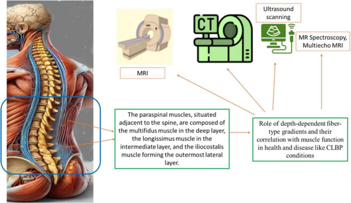
Methods: A literature review was executed to ascertain the databases including PubMed, Google Scholar, RelMed, and the National Library of Medicine. The search included studies elucidating recent imaging advancements, fiber-type composition analysis, level/depth-specific muscle characteristics, and clinical applications of novel radiological techniques in evaluating paraspinal muscle morphology and function. We performed this review without comprehensive meta-analysis.
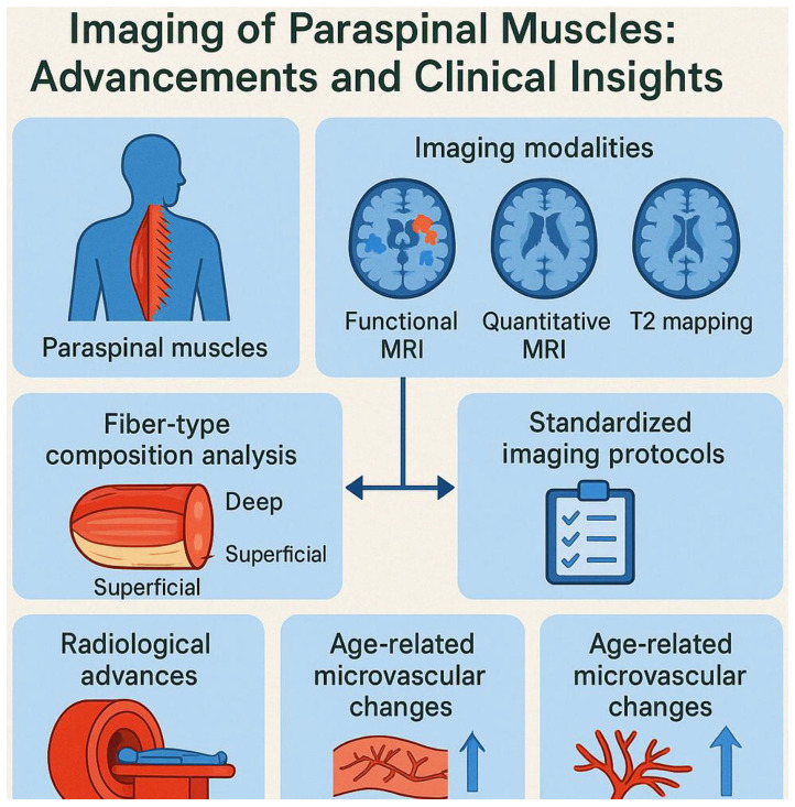
Results: The review identified significant advancements in imaging modalities for assessing paraspinal muscles, including functional MRI (fMRI), quantitative MRI (qMRI), and T2 mapping techniques. Key findings include: Fiber-type composition analysis: Recent studies elucidate the role of depth-dependent fiber-type gradients along with their correlation with muscle function in health and disease. Standardized imaging protocols: The lack of uniform imaging protocols remains a challenge, emphasizing the need for standardization to improve reproducibility and reliability. Radiological advances: Emerging techniques such as advanced fMRI and qMRI enable detailed visualization of muscle structure and function, overcoming limitations of traditional imaging methods. Age-related microvascular changes: age-related microvascular alterations significantly impact paraspinal muscle morphology and can be effectively captured by modern imaging biomarkers.
Conclusion: Advances in imaging techniques have enhanced our understanding of the structural and functional changes in paraspinal muscles associated with CLBP. The integration of imaging biomarkers into clinical practice holds promise for early diagnosis, targeted interventions, and better prognostic evaluations. Future research should focus on developing standardized imaging protocols and further exploring depth-specific properties of paraspinal muscles to enhance clinical outcomes.
期刊介绍:
Frontiers in Medicine publishes rigorously peer-reviewed research linking basic research to clinical practice and patient care, as well as translating scientific advances into new therapies and diagnostic tools. Led by an outstanding Editorial Board of international experts, this multidisciplinary open-access journal is at the forefront of disseminating and communicating scientific knowledge and impactful discoveries to researchers, academics, clinicians and the public worldwide.
In addition to papers that provide a link between basic research and clinical practice, a particular emphasis is given to studies that are directly relevant to patient care. In this spirit, the journal publishes the latest research results and medical knowledge that facilitate the translation of scientific advances into new therapies or diagnostic tools. The full listing of the Specialty Sections represented by Frontiers in Medicine is as listed below. As well as the established medical disciplines, Frontiers in Medicine is launching new sections that together will facilitate
- the use of patient-reported outcomes under real world conditions
- the exploitation of big data and the use of novel information and communication tools in the assessment of new medicines
- the scientific bases for guidelines and decisions from regulatory authorities
- access to medicinal products and medical devices worldwide
- addressing the grand health challenges around the world


 求助内容:
求助内容: 应助结果提醒方式:


