Renli Yang, Xirui Zhang, Yanjun Zhang, Yi Man, Xingmei Yang
{"title":"Low-Intensity Pulsed Ultrasound Promotes a Treg-Like Phenotype and Suppresses a Th17-Like Phenotype in CD4<sup>+</sup> T Cells via YAP/TAZ Activation in vitro.","authors":"Renli Yang, Xirui Zhang, Yanjun Zhang, Yi Man, Xingmei Yang","doi":"10.2147/JIR.S548291","DOIUrl":null,"url":null,"abstract":"<p><strong>Introduction: </strong>CD4<sup>+</sup> T cell subpopulations, particularly T helper 17 (Th17) and regulatory T (Treg) cells, exhibit antagonistic functions and play essential roles in inflammatory responses. Yes-associated protein (YAP) and transcriptional coactivator with PDZ-binding motif (TAZ) are critical modulators of cell proliferation and differentiation. Low-intensity pulsed ultrasound (LIPUS) has been shown to regulate YAP/TAZ activity, but its role in Th17/Treg balance remains unclear.</p><p><strong>Methods: </strong>CD4<sup>+</sup> T cells were purified from rat peripheral blood mononuclear cells (PBMCs) using magnetic-activated cell sorting (MACS). The cells were then treated with low-intensity pulsed ultrasound (LIPUS) at a set of parameters (1.0 MHz, 20 mW/cm², 20% duty cycle, 2h/day for 3 days) optimized based on preliminary proliferation and apoptosis assays. The effects of LIPUS on the expression of key functional markers (Foxp3 for Treg-like cells and IL-17A for Th17-like cells) were evaluated by flow cytometry, quantitative PCR, and ELISA. The activation and subcellular localization of YAP/TAZ were examined using immunofluorescence staining. Furthermore, siRNA-mediated knockdown was performed to investigate the functional involvement of YAP/TAZ in the LIPUS-mediated effects.</p><p><strong>Results: </strong>LIPUS treatment significantly increased the frequency of Foxp3-expressing cells while decreasing the frequency of IL-17A-producing cells. Additionally, LIPUS promoted the activation and nuclear translocation of YAP and TAZ, as evidenced by enhanced protein expression and a shift in subcellular localization. siRNA-mediated knockdown of YAP/TAZ attenuated the LIPUS-induced increase in Foxp3<sup>+</sup> cells and potentiated the population of IL-17A<sup>+</sup> cells. Importantly, LIPUS treatment effectively rescued the expression patterns of these functional markers following YAP/TAZ inhibition.</p><p><strong>Discussion: </strong>Our findings demonstrate that LIPUS promotes a Treg-like phenotype and suppresses a Th17-like phenotype in CD4<sup>+</sup> T cells, a process that is mediated, at least in part, by the activation of the YAP/TAZ signaling pathway. This immunomodulatory effect suggests that LIPUS could be explored as a novel non-invasive strategy for managing autoimmune diseases and chronic inflammatory conditions associated with an imbalance in T cell responses.</p>","PeriodicalId":16107,"journal":{"name":"Journal of Inflammation Research","volume":"18 ","pages":"13593-13608"},"PeriodicalIF":4.1000,"publicationDate":"2025-10-02","publicationTypes":"Journal Article","fieldsOfStudy":null,"isOpenAccess":false,"openAccessPdf":"https://www.ncbi.nlm.nih.gov/pmc/articles/PMC12499271/pdf/","citationCount":"0","resultStr":null,"platform":"Semanticscholar","paperid":null,"PeriodicalName":"Journal of Inflammation Research","FirstCategoryId":"3","ListUrlMain":"https://doi.org/10.2147/JIR.S548291","RegionNum":2,"RegionCategory":"医学","ArticlePicture":[],"TitleCN":null,"AbstractTextCN":null,"PMCID":null,"EPubDate":"2025/1/1 0:00:00","PubModel":"eCollection","JCR":"Q2","JCRName":"IMMUNOLOGY","Score":null,"Total":0}
引用次数: 0
Abstract
Introduction: CD4+ T cell subpopulations, particularly T helper 17 (Th17) and regulatory T (Treg) cells, exhibit antagonistic functions and play essential roles in inflammatory responses. Yes-associated protein (YAP) and transcriptional coactivator with PDZ-binding motif (TAZ) are critical modulators of cell proliferation and differentiation. Low-intensity pulsed ultrasound (LIPUS) has been shown to regulate YAP/TAZ activity, but its role in Th17/Treg balance remains unclear.
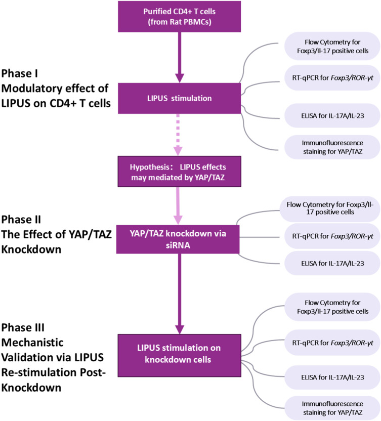
Methods: CD4+ T cells were purified from rat peripheral blood mononuclear cells (PBMCs) using magnetic-activated cell sorting (MACS). The cells were then treated with low-intensity pulsed ultrasound (LIPUS) at a set of parameters (1.0 MHz, 20 mW/cm², 20% duty cycle, 2h/day for 3 days) optimized based on preliminary proliferation and apoptosis assays. The effects of LIPUS on the expression of key functional markers (Foxp3 for Treg-like cells and IL-17A for Th17-like cells) were evaluated by flow cytometry, quantitative PCR, and ELISA. The activation and subcellular localization of YAP/TAZ were examined using immunofluorescence staining. Furthermore, siRNA-mediated knockdown was performed to investigate the functional involvement of YAP/TAZ in the LIPUS-mediated effects.
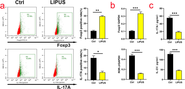
Results: LIPUS treatment significantly increased the frequency of Foxp3-expressing cells while decreasing the frequency of IL-17A-producing cells. Additionally, LIPUS promoted the activation and nuclear translocation of YAP and TAZ, as evidenced by enhanced protein expression and a shift in subcellular localization. siRNA-mediated knockdown of YAP/TAZ attenuated the LIPUS-induced increase in Foxp3+ cells and potentiated the population of IL-17A+ cells. Importantly, LIPUS treatment effectively rescued the expression patterns of these functional markers following YAP/TAZ inhibition.
Discussion: Our findings demonstrate that LIPUS promotes a Treg-like phenotype and suppresses a Th17-like phenotype in CD4+ T cells, a process that is mediated, at least in part, by the activation of the YAP/TAZ signaling pathway. This immunomodulatory effect suggests that LIPUS could be explored as a novel non-invasive strategy for managing autoimmune diseases and chronic inflammatory conditions associated with an imbalance in T cell responses.
期刊介绍:
An international, peer-reviewed, open access, online journal that welcomes laboratory and clinical findings on the molecular basis, cell biology and pharmacology of inflammation.

 求助内容:
求助内容: 应助结果提醒方式:


