Beatrice Longhi, Francesco Pera, Maria Menini, Francesco Bagnasco, Paolo Pesce, Marino Caroprese, Giuseppe Troiano, Khrystyna Zhurakivska
{"title":"The Influence of Implant Surface Modification on Marginal Bone Loss and Periodontal Health: A Cross-Over Randomized Clinical Trial.","authors":"Beatrice Longhi, Francesco Pera, Maria Menini, Francesco Bagnasco, Paolo Pesce, Marino Caroprese, Giuseppe Troiano, Khrystyna Zhurakivska","doi":"10.1155/ijod/8889144","DOIUrl":null,"url":null,"abstract":"<p><p><b>Background:</b> Edentulism rehabilitation with short implants is a procedure of proven efficacy. To improve the biological aspects of the interface between the implant and hard and soft tissues, different implant and prosthetic surface treatments have been proposed, producing contrasting results. The aim of present study is to compare implants and transmucosal components with an anodized collar with those with a traditionally machined collar in terms of Marginal bone loss and periodontal indexes. <b>Materials and Methods:</b> 30 patients were treated with two adjacent 6 mm length and 4.3 mm diameter implants (Shard short, Mech&Human, Grisignano di Zocco, Italy), one with an anodized collar (Test group) and one with a traditional machined collar (Control group), randomly positioned. Definitive transmucosal straight multiunit abutments (MUAs) (Mech&Human, Grisignano di Zocco, Italy) of height 1 mm, with differentially treated surfaces, were immediately screwed. After 3 months, prosthetic rehabilitation with splinted zirconia crowns screwed to the MUAs was made. Marginal bone levels (MBLs) were evaluated at the time of implant placement (T0), after 3 months (T3), after 6 and 12 months (T6 and T12) through periapical radiographies. Periodontal indexes (probing depth [PD], bleeding on probing [BoP], and plaque index [PlI]) were evaluated at the same timepoints, with the maximum follow-up of 12 months. <b>Results:</b> Average marginal bone loss at T3 was 0.40 ± 0.31 mm in the Test group and 0.42 ± 0.40 mm in the Control group (<i>p</i>=0.76), reaching 0.63 ± 0.41 and 0.78 ± 0.43 mm at T12 in the Test and the Control groups, respectively (<i>p</i>=0.94). Physiological PDs, with average values ranging between 1.48 and 2.1 mm, were revealed around the implants in both the groups, and The PlI ranged between 0 and 1 in most cases, and BoP appeared in some cases with isolated bleeding spots after probe passing (mean values ranging between 0.20 ± 0.41 and 0.50 ± 0.52), with no significant differences between groups. <b>Conclusions:</b> Surface treatment with anodization of implant collar and transmucosal components seem to not influence marginal bone stability at 1-year follow-up, nor the condition of periodontal tissues. Long-term follow-ups are needed to confirm the results. <b>Trial Registration</b>: ClinicalTrials.gov identifier: NCT05766878.</p>","PeriodicalId":13947,"journal":{"name":"International Journal of Dentistry","volume":"2025 ","pages":"8889144"},"PeriodicalIF":2.2000,"publicationDate":"2025-09-28","publicationTypes":"Journal Article","fieldsOfStudy":null,"isOpenAccess":false,"openAccessPdf":"https://www.ncbi.nlm.nih.gov/pmc/articles/PMC12497520/pdf/","citationCount":"0","resultStr":null,"platform":"Semanticscholar","paperid":null,"PeriodicalName":"International Journal of Dentistry","FirstCategoryId":"1085","ListUrlMain":"https://doi.org/10.1155/ijod/8889144","RegionNum":0,"RegionCategory":null,"ArticlePicture":[],"TitleCN":null,"AbstractTextCN":null,"PMCID":null,"EPubDate":"2025/1/1 0:00:00","PubModel":"eCollection","JCR":"Q2","JCRName":"DENTISTRY, ORAL SURGERY & MEDICINE","Score":null,"Total":0}
引用次数: 0
Abstract
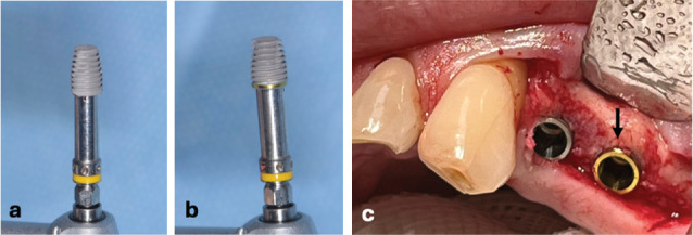
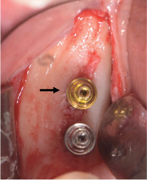
Background: Edentulism rehabilitation with short implants is a procedure of proven efficacy. To improve the biological aspects of the interface between the implant and hard and soft tissues, different implant and prosthetic surface treatments have been proposed, producing contrasting results. The aim of present study is to compare implants and transmucosal components with an anodized collar with those with a traditionally machined collar in terms of Marginal bone loss and periodontal indexes. Materials and Methods: 30 patients were treated with two adjacent 6 mm length and 4.3 mm diameter implants (Shard short, Mech&Human, Grisignano di Zocco, Italy), one with an anodized collar (Test group) and one with a traditional machined collar (Control group), randomly positioned. Definitive transmucosal straight multiunit abutments (MUAs) (Mech&Human, Grisignano di Zocco, Italy) of height 1 mm, with differentially treated surfaces, were immediately screwed. After 3 months, prosthetic rehabilitation with splinted zirconia crowns screwed to the MUAs was made. Marginal bone levels (MBLs) were evaluated at the time of implant placement (T0), after 3 months (T3), after 6 and 12 months (T6 and T12) through periapical radiographies. Periodontal indexes (probing depth [PD], bleeding on probing [BoP], and plaque index [PlI]) were evaluated at the same timepoints, with the maximum follow-up of 12 months. Results: Average marginal bone loss at T3 was 0.40 ± 0.31 mm in the Test group and 0.42 ± 0.40 mm in the Control group (p=0.76), reaching 0.63 ± 0.41 and 0.78 ± 0.43 mm at T12 in the Test and the Control groups, respectively (p=0.94). Physiological PDs, with average values ranging between 1.48 and 2.1 mm, were revealed around the implants in both the groups, and The PlI ranged between 0 and 1 in most cases, and BoP appeared in some cases with isolated bleeding spots after probe passing (mean values ranging between 0.20 ± 0.41 and 0.50 ± 0.52), with no significant differences between groups. Conclusions: Surface treatment with anodization of implant collar and transmucosal components seem to not influence marginal bone stability at 1-year follow-up, nor the condition of periodontal tissues. Long-term follow-ups are needed to confirm the results. Trial Registration: ClinicalTrials.gov identifier: NCT05766878.

 求助内容:
求助内容: 应助结果提醒方式:


