TP53 expression in PCAT in coronary artery disease combined with type 2 diabetes mellitus and its correlation with CCTA radiomic features: novel imaging biomarkers.
Haicheng Qi, Yahui Hu, Yan Li, Xiumei Li, Yan Xing
{"title":"<i>TP53</i> expression in PCAT in coronary artery disease combined with type 2 diabetes mellitus and its correlation with CCTA radiomic features: novel imaging biomarkers.","authors":"Haicheng Qi, Yahui Hu, Yan Li, Xiumei Li, Yan Xing","doi":"10.3389/fmed.2025.1626390","DOIUrl":null,"url":null,"abstract":"<p><strong>Aims: </strong>To explore the changes in differentially expressed genes in pericoronary adipose tissue (PCAT) and serum from patients with coronary artery disease (CAD) complicated with type 2 diabetes mellitus (T2DM) and to analyse its correlation with PCAT radiomic features based on coronary CT angiography (CCTA).</p><p><strong>Methods: </strong>Intersecting genes that were differentially expressed in both CAD and T2DM patients were obtained from the GEO database and analyzed to obtain candidate genes. PCAT and serum samples were collected from CAD patients who underwent coronary artery bypass grafting (CABG) from May 2023 to January 2024. RT-qPCR was used to determine the expression of candidate differentially expressed genes in PCAT, to search for genes related to patients with CAD combined with T2DM, and to verify the protein expression levels by immunohistochemistry (IHC). Enzyme-linked immunosorbent assays (ELISAs) were also used to determine the expression of candidate differentially expressed genes in the serum. Finally, the PCAT radiomic features of the right coronary artery in patients with CAD combined with T2DM were extracted and correlated with the candidate genes.</p><p><strong>Results: </strong><i>HLA-DRB1</i>, <i>TP53</i>, and <i>CCR9</i> were screened from the GEO database. RT-qPCR results revealed that <i>TP53</i> expression was significantly increased in the T2DM group compared with the control group (3.082 ± 0.580 vs. 1.663 ± 0.698, <i>p</i> < 0.001). IHC results revealed that the area of positive expression around the nucleus was increased in the fat cells of the T2DM group compared with those of the control group, with increased perinuclear areas with positive expression (0.521 ± 0.082 vs. 0.327 ± 0.074, <i>p</i> < 0.001), and 14 PCAT radiomic features in CAD combined with T2DM patients correlated with <i>TP53</i> (r<sub>s</sub> > 0.5, <i>p</i> < 0.05).</p><p><strong>Conclusion: </strong><i>TP53</i> expression was significantly elevated in the PCAT of patients with CAD combined with T2DM, suggesting that this molecule plays a role in the development of this disease. Four first-order features and 10 texture features in the PCAT radiomic features were associated with abnormal <i>TP53</i> expression. The association of radiomic features with <i>TP53</i> suggests that CCTA-based radiomic features can be used to predict abnormalities in differential gene expression, thus providing a new way to noninvasively predict CAD combined with T2DM.</p>","PeriodicalId":12488,"journal":{"name":"Frontiers in Medicine","volume":"12 ","pages":"1626390"},"PeriodicalIF":3.1000,"publicationDate":"2025-09-22","publicationTypes":"Journal Article","fieldsOfStudy":null,"isOpenAccess":false,"openAccessPdf":"https://www.ncbi.nlm.nih.gov/pmc/articles/PMC12497776/pdf/","citationCount":"0","resultStr":null,"platform":"Semanticscholar","paperid":null,"PeriodicalName":"Frontiers in Medicine","FirstCategoryId":"3","ListUrlMain":"https://doi.org/10.3389/fmed.2025.1626390","RegionNum":3,"RegionCategory":"医学","ArticlePicture":[],"TitleCN":null,"AbstractTextCN":null,"PMCID":null,"EPubDate":"2025/1/1 0:00:00","PubModel":"eCollection","JCR":"Q1","JCRName":"MEDICINE, GENERAL & INTERNAL","Score":null,"Total":0}
引用次数: 0
Abstract
Aims: To explore the changes in differentially expressed genes in pericoronary adipose tissue (PCAT) and serum from patients with coronary artery disease (CAD) complicated with type 2 diabetes mellitus (T2DM) and to analyse its correlation with PCAT radiomic features based on coronary CT angiography (CCTA).
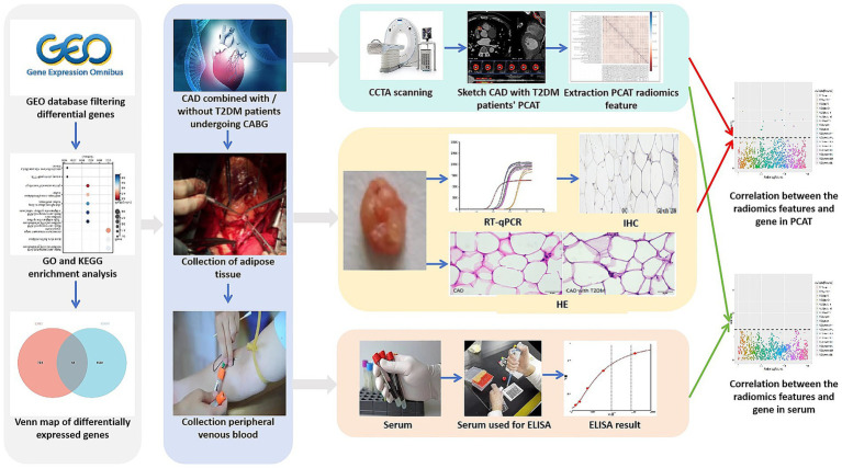
Methods: Intersecting genes that were differentially expressed in both CAD and T2DM patients were obtained from the GEO database and analyzed to obtain candidate genes. PCAT and serum samples were collected from CAD patients who underwent coronary artery bypass grafting (CABG) from May 2023 to January 2024. RT-qPCR was used to determine the expression of candidate differentially expressed genes in PCAT, to search for genes related to patients with CAD combined with T2DM, and to verify the protein expression levels by immunohistochemistry (IHC). Enzyme-linked immunosorbent assays (ELISAs) were also used to determine the expression of candidate differentially expressed genes in the serum. Finally, the PCAT radiomic features of the right coronary artery in patients with CAD combined with T2DM were extracted and correlated with the candidate genes.
Results: HLA-DRB1, TP53, and CCR9 were screened from the GEO database. RT-qPCR results revealed that TP53 expression was significantly increased in the T2DM group compared with the control group (3.082 ± 0.580 vs. 1.663 ± 0.698, p < 0.001). IHC results revealed that the area of positive expression around the nucleus was increased in the fat cells of the T2DM group compared with those of the control group, with increased perinuclear areas with positive expression (0.521 ± 0.082 vs. 0.327 ± 0.074, p < 0.001), and 14 PCAT radiomic features in CAD combined with T2DM patients correlated with TP53 (rs > 0.5, p < 0.05).
Conclusion: TP53 expression was significantly elevated in the PCAT of patients with CAD combined with T2DM, suggesting that this molecule plays a role in the development of this disease. Four first-order features and 10 texture features in the PCAT radiomic features were associated with abnormal TP53 expression. The association of radiomic features with TP53 suggests that CCTA-based radiomic features can be used to predict abnormalities in differential gene expression, thus providing a new way to noninvasively predict CAD combined with T2DM.
期刊介绍:
Frontiers in Medicine publishes rigorously peer-reviewed research linking basic research to clinical practice and patient care, as well as translating scientific advances into new therapies and diagnostic tools. Led by an outstanding Editorial Board of international experts, this multidisciplinary open-access journal is at the forefront of disseminating and communicating scientific knowledge and impactful discoveries to researchers, academics, clinicians and the public worldwide.
In addition to papers that provide a link between basic research and clinical practice, a particular emphasis is given to studies that are directly relevant to patient care. In this spirit, the journal publishes the latest research results and medical knowledge that facilitate the translation of scientific advances into new therapies or diagnostic tools. The full listing of the Specialty Sections represented by Frontiers in Medicine is as listed below. As well as the established medical disciplines, Frontiers in Medicine is launching new sections that together will facilitate
- the use of patient-reported outcomes under real world conditions
- the exploitation of big data and the use of novel information and communication tools in the assessment of new medicines
- the scientific bases for guidelines and decisions from regulatory authorities
- access to medicinal products and medical devices worldwide
- addressing the grand health challenges around the world


 求助内容:
求助内容: 应助结果提醒方式:


