Influence of prone, supine, and lateral positions during spine surgery on vascular, abdominal, and postural anatomy: a comprehensive review and Bayesian meta-analysis.
{"title":"Influence of prone, supine, and lateral positions during spine surgery on vascular, abdominal, and postural anatomy: a comprehensive review and Bayesian meta-analysis.","authors":"Samir Smajic, Markus Konieczny, Koroush Kabir, Raffaele Scrofani, Filippo Migliorini, Anel Dracic","doi":"10.1186/s40001-025-03239-2","DOIUrl":null,"url":null,"abstract":"<p><strong>Background: </strong>Patient positioning alters the three-dimensional relationship between the spine and surrounding neurovascular and visceral structures, thereby influencing both the technical feasibility and safety of lumbar procedures. Quantitative estimates of these positional shifts remain heterogeneous.</p><p><strong>Objective: </strong>To determine, across contemporary imaging studies, how prone, supine, and lateral decubitus positions alter the displacement of great vessels and retroperitoneal organs, the location of the psoas/lumbar plexus, and segmental lumbar lordosis.</p><p><strong>Methods: </strong>MEDLINE, Embase, and CENTRAL were searched from 2015 to 2025. Eligible studies compared at least two positions in adults and reported millimetre or degree differences for the outcomes of interest. Random‑effects (REML) subgroup meta‑analyses, a graph‑theoretical network meta‑analysis (netmeta), leave‑one‑out diagnostics, and Bayesian sensitivity models were performed. Risk of bias was assessed with ROBINS‑I.</p><p><strong>Results: </strong>Nine studies (41 independent comparisons; n = 1,248) met inclusion criteria. Retro‑peritoneal organs moved posteriorly by a pooled + 6.34 mm (95% CI 1.87-10.80; p = 0.007) when patients were turned from lateral decubitus to the prone position, narrowing the anterior working corridor at L2-L4. No significant pooled displacement was detected for major vessels (+ 1.26 mm, 95% CI -2.43-4.94), psoas/plexus (+ 0.94 mm, 95% CI -3.58-5.46) or segmental lordosis (+ 1.55°, 95% CI -4.62-7.73°). Direct contrasts showed that the supine-to-prone transition increased combined displacement/lordosis by + 3.64 mm / °(95% CI 0.53-6.76). Network ranking favoured the supine position for anatomical stability, but inconsistency was high (I<sup>2</sup> = 89%). Two studies were low, three moderate, three serious and one critical risk of bias; removing serious/critical studies did not change the effect direction.</p><p><strong>Conclusions: </strong>Turning a patient prone produces a reproducible posterior migration of the colon and kidney (6 mm) and a modest increase in lumbar lordosis (3-4°). Vascular and psoas positions are highly patient-specific and cannot be assumed based on supine imaging alone. Preoperative planning should therefore incorporate position-matched imaging or intraoperative navigation, especially for anterior or anterolateral approaches at L2-L4. Further high-quality, multi-positional imaging studies are warranted to clarify the sources of the marked heterogeneity observed.</p>","PeriodicalId":11949,"journal":{"name":"European Journal of Medical Research","volume":"30 1","pages":"932"},"PeriodicalIF":3.4000,"publicationDate":"2025-10-07","publicationTypes":"Journal Article","fieldsOfStudy":null,"isOpenAccess":false,"openAccessPdf":"https://www.ncbi.nlm.nih.gov/pmc/articles/PMC12502251/pdf/","citationCount":"0","resultStr":null,"platform":"Semanticscholar","paperid":null,"PeriodicalName":"European Journal of Medical Research","FirstCategoryId":"3","ListUrlMain":"https://doi.org/10.1186/s40001-025-03239-2","RegionNum":3,"RegionCategory":"医学","ArticlePicture":[],"TitleCN":null,"AbstractTextCN":null,"PMCID":null,"EPubDate":"","PubModel":"","JCR":"Q2","JCRName":"MEDICINE, RESEARCH & EXPERIMENTAL","Score":null,"Total":0}
引用次数: 0
Abstract
Background: Patient positioning alters the three-dimensional relationship between the spine and surrounding neurovascular and visceral structures, thereby influencing both the technical feasibility and safety of lumbar procedures. Quantitative estimates of these positional shifts remain heterogeneous.
Objective: To determine, across contemporary imaging studies, how prone, supine, and lateral decubitus positions alter the displacement of great vessels and retroperitoneal organs, the location of the psoas/lumbar plexus, and segmental lumbar lordosis.
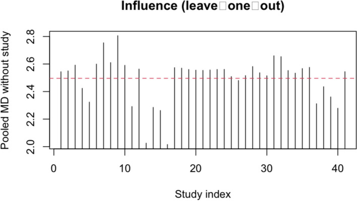
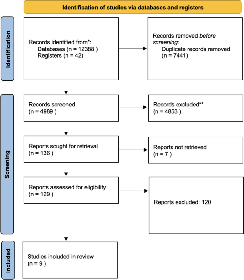
Methods: MEDLINE, Embase, and CENTRAL were searched from 2015 to 2025. Eligible studies compared at least two positions in adults and reported millimetre or degree differences for the outcomes of interest. Random‑effects (REML) subgroup meta‑analyses, a graph‑theoretical network meta‑analysis (netmeta), leave‑one‑out diagnostics, and Bayesian sensitivity models were performed. Risk of bias was assessed with ROBINS‑I.
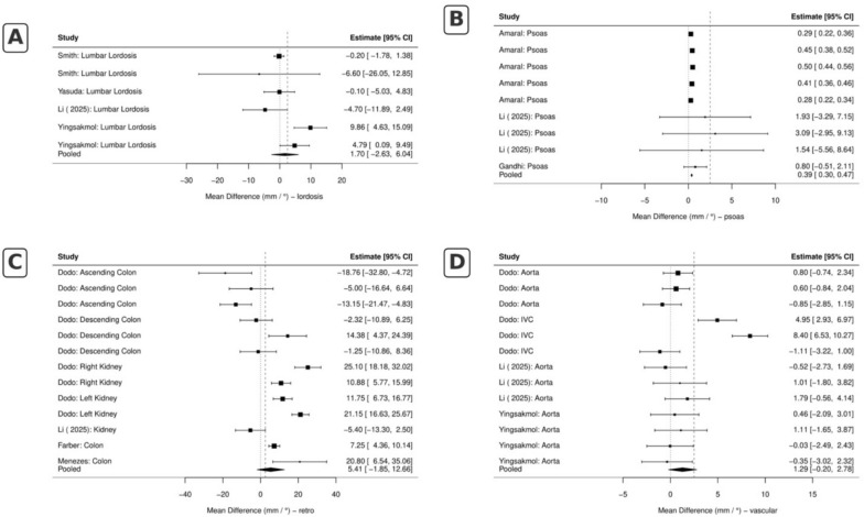
Results: Nine studies (41 independent comparisons; n = 1,248) met inclusion criteria. Retro‑peritoneal organs moved posteriorly by a pooled + 6.34 mm (95% CI 1.87-10.80; p = 0.007) when patients were turned from lateral decubitus to the prone position, narrowing the anterior working corridor at L2-L4. No significant pooled displacement was detected for major vessels (+ 1.26 mm, 95% CI -2.43-4.94), psoas/plexus (+ 0.94 mm, 95% CI -3.58-5.46) or segmental lordosis (+ 1.55°, 95% CI -4.62-7.73°). Direct contrasts showed that the supine-to-prone transition increased combined displacement/lordosis by + 3.64 mm / °(95% CI 0.53-6.76). Network ranking favoured the supine position for anatomical stability, but inconsistency was high (I2 = 89%). Two studies were low, three moderate, three serious and one critical risk of bias; removing serious/critical studies did not change the effect direction.
Conclusions: Turning a patient prone produces a reproducible posterior migration of the colon and kidney (6 mm) and a modest increase in lumbar lordosis (3-4°). Vascular and psoas positions are highly patient-specific and cannot be assumed based on supine imaging alone. Preoperative planning should therefore incorporate position-matched imaging or intraoperative navigation, especially for anterior or anterolateral approaches at L2-L4. Further high-quality, multi-positional imaging studies are warranted to clarify the sources of the marked heterogeneity observed.
背景:患者体位改变了脊柱与周围神经血管和内脏结构之间的三维关系,从而影响腰椎手术的技术可行性和安全性。对这些位置变化的定量估计仍然不一致。目的:通过当代影像学研究,确定俯卧位、仰卧位和侧卧位如何改变大血管和腹膜后器官的移位、腰肌/腰丛的位置和腰椎前凸。方法:检索2015 - 2025年MEDLINE、Embase和CENTRAL数据库。符合条件的研究比较了至少两种成人体位,并报告了感兴趣的结果的毫米或程度差异。进行了随机效应(REML)亚组元分析、图理论网络元分析(netmeta)、遗漏诊断和贝叶斯灵敏度模型。采用ROBINS - I评估偏倚风险。结果:9项研究(41项独立比较,n = 1,248)符合纳入标准。当患者从侧卧位转到俯卧位时,腹膜脏器向后移动了6.34 mm(95% CI 1.87-10.80;p = 0.007),使L2-L4的前工作通道变窄。主要血管(+ 1.26 mm, 95% CI -2.43-4.94)、腰肌/神经丛(+ 0.94 mm, 95% CI -3.58-5.46)或节段性前凸(+ 1.55°,95% CI -4.62-7.73°)均未检测到明显的合并移位。直接对比显示,仰卧位到俯卧位的转变使联合位移/前凸增加了+ 3.64 mm / °(95% CI 0.53-6.76)。网络排序有利于仰卧位的解剖稳定性,但不一致性很高(I2 = 89%)。2项研究为低偏倚风险,3项为中度偏倚风险,3项为严重偏倚风险,1项为严重偏倚风险;删除严肃/关键的研究并没有改变效应的方向。结论:使患者俯卧可产生可重复的结肠和肾脏后移(6mm),腰椎前凸轻度增加(3-4°)。血管和腰肌的位置是高度患者特异性的,不能仅根据仰卧位成像来假设。因此,术前规划应结合位置匹配成像或术中导航,特别是L2-L4的前路或前外侧入路。进一步的高质量、多位置成像研究是必要的,以澄清观察到的明显异质性的来源。
期刊介绍:
European Journal of Medical Research publishes translational and clinical research of international interest across all medical disciplines, enabling clinicians and other researchers to learn about developments and innovations within these disciplines and across the boundaries between disciplines. The journal publishes high quality research and reviews and aims to ensure that the results of all well-conducted research are published, regardless of their outcome.
 求助内容:
求助内容: 应助结果提醒方式:


