Geeta Lalwani, Charles C Wykoff, Jayla Briggs, Chin-Yu Lin, Verena Steffen, Zdenka Haskova
{"title":"Progression of diabetic retinopathy in a longitudinal real-world study of patients in primary care.","authors":"Geeta Lalwani, Charles C Wykoff, Jayla Briggs, Chin-Yu Lin, Verena Steffen, Zdenka Haskova","doi":"10.1186/s12886-025-04307-1","DOIUrl":null,"url":null,"abstract":"<p><strong>Background: </strong>The aim of this study was to assess the impact of diabetic retinopathy (DR) severity on the risk of DR progression to proliferative DR (PDR) or clinically significant macular edema (CSME) in patients with diabetes mellitus (DM).</p><p><strong>Methods: </strong>This is a prospective, longitudinal, non-interventional, and observational cohort study of patients with DM in the United States based on a database of 7-field color fundus photograph images from primary care visits. Study participants were adults aged ≥ 18 years who underwent DR screening in either eye between January 1999 and December 2016. DR severity was graded according to the Early Treatment Diabetic Retinopathy Study-Diabetic Retinopathy Severity Scale (ETDRS-DRSS) on 7-field color fundus photographs. The main outcomes were ≥ 2-step DR worsening and development of CSME, PDR, or CSME and PDR together.</p><p><strong>Results: </strong>For all 41,977 eyes evaluated, the proportion of eyes with ≥ 2-step DR worsening was 2.7% at year 2 and 9.6% at year 5. Rate of ≥ 2-step DR worsening was greatest among eyes with moderate-to-severe NPDR with baseline DRSS 43-53 (36.5% at year 5). Analysis of PDR and CSME outcomes showed the presence of 3 distinct clinical phenotypes: 1 subset progressed to CSME (1.24% at year 5), another to PDR (0.49% at year 5), and only a small subset progressed to both vision-threatening forms of DR (0.10% at year 5). The clinical phenotype did not appear to be dependent on baseline DRSS.</p><p><strong>Conclusions: </strong>Overall, the risk of progression to vision-threatening forms of DR was more pronounced in eyes with moderate-to-severe non-proliferative DR at baseline. In addition, we found that distinct DR clinical subtypes progressing to either PDR and/or CSME over a 5-year period are not driven by baseline DR severity, suggesting other factors may contribute.</p>","PeriodicalId":9058,"journal":{"name":"BMC Ophthalmology","volume":"25 1","pages":"547"},"PeriodicalIF":1.7000,"publicationDate":"2025-10-06","publicationTypes":"Journal Article","fieldsOfStudy":null,"isOpenAccess":false,"openAccessPdf":"https://www.ncbi.nlm.nih.gov/pmc/articles/PMC12502510/pdf/","citationCount":"0","resultStr":null,"platform":"Semanticscholar","paperid":null,"PeriodicalName":"BMC Ophthalmology","FirstCategoryId":"3","ListUrlMain":"https://doi.org/10.1186/s12886-025-04307-1","RegionNum":4,"RegionCategory":"医学","ArticlePicture":[],"TitleCN":null,"AbstractTextCN":null,"PMCID":null,"EPubDate":"","PubModel":"","JCR":"Q3","JCRName":"OPHTHALMOLOGY","Score":null,"Total":0}
引用次数: 0
Abstract
Background: The aim of this study was to assess the impact of diabetic retinopathy (DR) severity on the risk of DR progression to proliferative DR (PDR) or clinically significant macular edema (CSME) in patients with diabetes mellitus (DM).
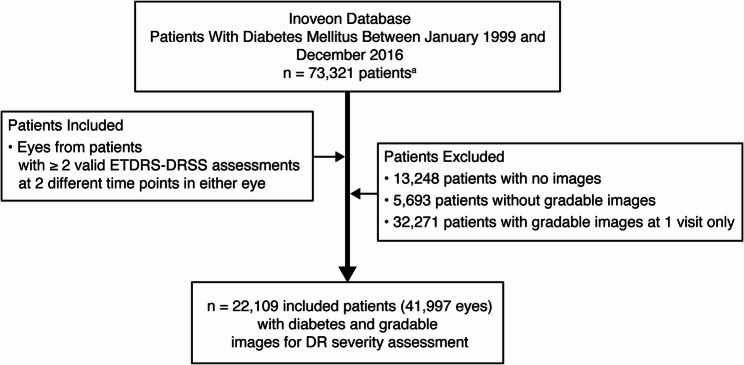
Methods: This is a prospective, longitudinal, non-interventional, and observational cohort study of patients with DM in the United States based on a database of 7-field color fundus photograph images from primary care visits. Study participants were adults aged ≥ 18 years who underwent DR screening in either eye between January 1999 and December 2016. DR severity was graded according to the Early Treatment Diabetic Retinopathy Study-Diabetic Retinopathy Severity Scale (ETDRS-DRSS) on 7-field color fundus photographs. The main outcomes were ≥ 2-step DR worsening and development of CSME, PDR, or CSME and PDR together.
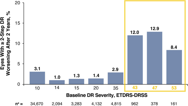
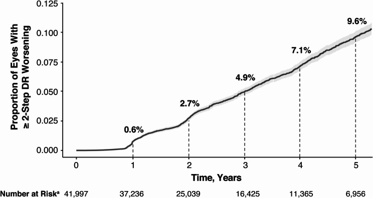
Results: For all 41,977 eyes evaluated, the proportion of eyes with ≥ 2-step DR worsening was 2.7% at year 2 and 9.6% at year 5. Rate of ≥ 2-step DR worsening was greatest among eyes with moderate-to-severe NPDR with baseline DRSS 43-53 (36.5% at year 5). Analysis of PDR and CSME outcomes showed the presence of 3 distinct clinical phenotypes: 1 subset progressed to CSME (1.24% at year 5), another to PDR (0.49% at year 5), and only a small subset progressed to both vision-threatening forms of DR (0.10% at year 5). The clinical phenotype did not appear to be dependent on baseline DRSS.
Conclusions: Overall, the risk of progression to vision-threatening forms of DR was more pronounced in eyes with moderate-to-severe non-proliferative DR at baseline. In addition, we found that distinct DR clinical subtypes progressing to either PDR and/or CSME over a 5-year period are not driven by baseline DR severity, suggesting other factors may contribute.
期刊介绍:
BMC Ophthalmology is an open access, peer-reviewed journal that considers articles on all aspects of the prevention, diagnosis and management of eye disorders, as well as related molecular genetics, pathophysiology, and epidemiology.

 求助内容:
求助内容: 应助结果提醒方式:


