Rongrong Miao, Tingting Wu, Qilong Wu, Teng Ye, Hui Li, Shusheng Liao
{"title":"Noninvasive assessment of hemodynamic profile and myocardial mechanics in pulsus alternans patients by multiple echocardiographic methods.","authors":"Rongrong Miao, Tingting Wu, Qilong Wu, Teng Ye, Hui Li, Shusheng Liao","doi":"10.1186/s13089-025-00448-y","DOIUrl":null,"url":null,"abstract":"<p><strong>Background: </strong>Pulsus alternans (PA) is an intriguing phenomenon and a clinically rare entity. Accurately assessing cardiac function in patients with PA remains challenging. This study aims to investigate the myocardial mechanical characteristics and non-invasive hemodynamic profiles of PA patients using multiple echocardiographic imaging modalities.</p><p><strong>Methods: </strong>Clinical and echocardiographic data were retrospectively analysed from 16 patients diagnosed with PA by echocardiography at our hospital between January 2021 and May 2025. In this study, the characteristics of PA were elaborated by multiple echocardiographic methods, and the non-invasive hemodynamic profile was determined by pulse-wave Doppler.</p><p><strong>Results: </strong>Sixteen patients were enrolled. Seven were classified as NYHA class III and six as class IV. Elevated levels of NT-proBNP and hs-cTNT were observed in most patients. Follow-up ranged from 1 to 44 months, and five patients experienced adverse outcomes, including heart transplantation, rehospitalisation, and death. Within this cohort, three patients exhibited biventricular PA, while 13 patients presented with left ventricular (LV) PA. Key hemodynamic parameters varied significantly: LVOT-VTI<sub>strong beat</sub> ranged from 11.3 cm to 29.2 cm, LVOT-VTI<sub>weak beat</sub> from 6.8 cm to 22.1 cm, and the variation rate between strong and weak beats (∆LVOT-VTI) ranged from 19 to 52%. Global longitudinal strain (GLS) was significantly reduced in 14 patients (range: - 1.2% to - 10.4%), while peak strain dispersion (PSD) increased (range: 47 ms to 117.5 ms). Two patients were excluded from strain analysis due to suboptimal imaging. Hemodynamic parameters (LVOT-VTI<sub>strong beat</sub>, LVOT-VTI<sub>weak beat</sub> and ∆LVOT-VTI) showed strong correlations with GLS in PA patients (r = 0.806, P = 0.001; r = 0.642, P = 0.018 and r = 0.611, P = 0.027, respectively). NT-proBNP was significantly positively related to adverse outcomes in PA patients (r = 0.669, P = 0.012).</p><p><strong>Conclusion: </strong>Echocardiography is essential for evaluating cardiac function in patients with PA. This study used multiple echocardiographic methods to delineate the characteristics of this intriguing clinical phenomenon. Non-invasive hemodynamic parameters are potentially important for prognosis assessment, and myocardial strain assessment provides valuable insights into myocardial mechanical features. A comprehensive analysis using multimodality imaging is crucial for accurately identifying this disease, potentially enhancing the understanding of the pathophysiological mechanism of PA.</p>","PeriodicalId":36911,"journal":{"name":"Ultrasound Journal","volume":"17 1","pages":"46"},"PeriodicalIF":2.9000,"publicationDate":"2025-10-06","publicationTypes":"Journal Article","fieldsOfStudy":null,"isOpenAccess":false,"openAccessPdf":"https://www.ncbi.nlm.nih.gov/pmc/articles/PMC12500491/pdf/","citationCount":"0","resultStr":null,"platform":"Semanticscholar","paperid":null,"PeriodicalName":"Ultrasound Journal","FirstCategoryId":"1085","ListUrlMain":"https://doi.org/10.1186/s13089-025-00448-y","RegionNum":0,"RegionCategory":null,"ArticlePicture":[],"TitleCN":null,"AbstractTextCN":null,"PMCID":null,"EPubDate":"","PubModel":"","JCR":"Q2","JCRName":"Medicine","Score":null,"Total":0}
引用次数: 0
Abstract
Background: Pulsus alternans (PA) is an intriguing phenomenon and a clinically rare entity. Accurately assessing cardiac function in patients with PA remains challenging. This study aims to investigate the myocardial mechanical characteristics and non-invasive hemodynamic profiles of PA patients using multiple echocardiographic imaging modalities.
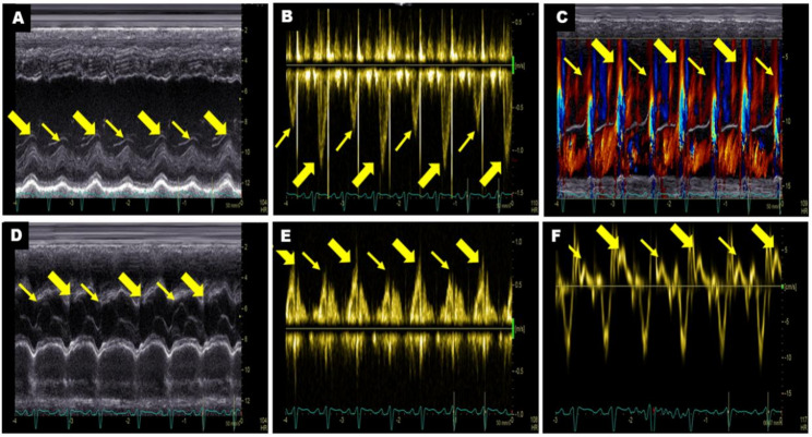
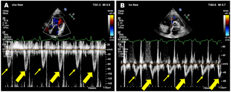
Methods: Clinical and echocardiographic data were retrospectively analysed from 16 patients diagnosed with PA by echocardiography at our hospital between January 2021 and May 2025. In this study, the characteristics of PA were elaborated by multiple echocardiographic methods, and the non-invasive hemodynamic profile was determined by pulse-wave Doppler.
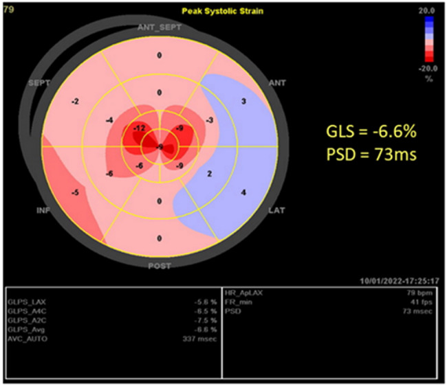
Results: Sixteen patients were enrolled. Seven were classified as NYHA class III and six as class IV. Elevated levels of NT-proBNP and hs-cTNT were observed in most patients. Follow-up ranged from 1 to 44 months, and five patients experienced adverse outcomes, including heart transplantation, rehospitalisation, and death. Within this cohort, three patients exhibited biventricular PA, while 13 patients presented with left ventricular (LV) PA. Key hemodynamic parameters varied significantly: LVOT-VTIstrong beat ranged from 11.3 cm to 29.2 cm, LVOT-VTIweak beat from 6.8 cm to 22.1 cm, and the variation rate between strong and weak beats (∆LVOT-VTI) ranged from 19 to 52%. Global longitudinal strain (GLS) was significantly reduced in 14 patients (range: - 1.2% to - 10.4%), while peak strain dispersion (PSD) increased (range: 47 ms to 117.5 ms). Two patients were excluded from strain analysis due to suboptimal imaging. Hemodynamic parameters (LVOT-VTIstrong beat, LVOT-VTIweak beat and ∆LVOT-VTI) showed strong correlations with GLS in PA patients (r = 0.806, P = 0.001; r = 0.642, P = 0.018 and r = 0.611, P = 0.027, respectively). NT-proBNP was significantly positively related to adverse outcomes in PA patients (r = 0.669, P = 0.012).
Conclusion: Echocardiography is essential for evaluating cardiac function in patients with PA. This study used multiple echocardiographic methods to delineate the characteristics of this intriguing clinical phenomenon. Non-invasive hemodynamic parameters are potentially important for prognosis assessment, and myocardial strain assessment provides valuable insights into myocardial mechanical features. A comprehensive analysis using multimodality imaging is crucial for accurately identifying this disease, potentially enhancing the understanding of the pathophysiological mechanism of PA.

 求助内容:
求助内容: 应助结果提醒方式:


