{"title":"Machine learning-based assessment of condylar changes after orthognathic surgery of asymmetric dentofacial deformities.","authors":"Jin Tang, Jiayu Shi, Shuguang Liu","doi":"10.3389/fsurg.2025.1594849","DOIUrl":null,"url":null,"abstract":"<p><strong>Introduction: </strong>This retrospective study aimed to investigate three-dimensional (3D) condyle change including volumetric, positional, rotational alterations in patients with asymmetric dentofacial deformities immediate and long after bimaxillary orthognathic surgery.</p><p><strong>Methods: </strong>The sample included 36 patients who underwent bimaxillary orthognathic surgery, with a maxillomandibular dental midline deviation >3 mm, excluding those with class II/III malocclusions and craniofacial syndrome. A fully automated deep learning-based assessment method was used to analyze the volume, position and rotation of condyle based on Cone-beam Computed Tomography (CBCT) images. Repeated measures ANOVA was used to compared the changes at three intervals-pre-surgery (T0), one-week post-surgery (T1), and six months post-surgery (T2)-of the deviated sides and non-deviated sides condyle.</p><p><strong>Results: </strong>The condyle on the deviated side was smaller than that on the non-deviated side, with significant volume reductions observed six months post-surgery on the deviated side. Several condylar changes were observed immediately after surgery, though of small magnitude, and it mostly tended to return to their original positions 6 months after surgery. However, the condyle rotated laterally on the deviated side and medially on the non-deviated side post operation and in long-term.</p><p><strong>Conclusion: </strong>For patients with asymmetry, condyle on the deviated side undergo greater remodeling than the non-deviated side after orthognathic surgery. There are measurable rotations in the coronal plane of condyle on both sides.</p>","PeriodicalId":12564,"journal":{"name":"Frontiers in Surgery","volume":"12 ","pages":"1594849"},"PeriodicalIF":1.6000,"publicationDate":"2025-09-18","publicationTypes":"Journal Article","fieldsOfStudy":null,"isOpenAccess":false,"openAccessPdf":"https://www.ncbi.nlm.nih.gov/pmc/articles/PMC12488556/pdf/","citationCount":"0","resultStr":null,"platform":"Semanticscholar","paperid":null,"PeriodicalName":"Frontiers in Surgery","FirstCategoryId":"3","ListUrlMain":"https://doi.org/10.3389/fsurg.2025.1594849","RegionNum":4,"RegionCategory":"医学","ArticlePicture":[],"TitleCN":null,"AbstractTextCN":null,"PMCID":null,"EPubDate":"2025/1/1 0:00:00","PubModel":"eCollection","JCR":"Q2","JCRName":"SURGERY","Score":null,"Total":0}
引用次数: 0
Abstract
Introduction: This retrospective study aimed to investigate three-dimensional (3D) condyle change including volumetric, positional, rotational alterations in patients with asymmetric dentofacial deformities immediate and long after bimaxillary orthognathic surgery.
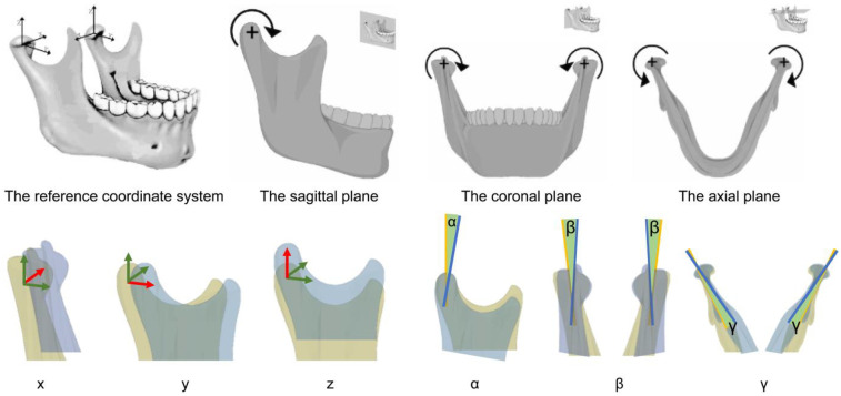
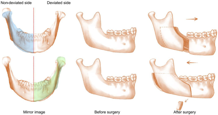
Methods: The sample included 36 patients who underwent bimaxillary orthognathic surgery, with a maxillomandibular dental midline deviation >3 mm, excluding those with class II/III malocclusions and craniofacial syndrome. A fully automated deep learning-based assessment method was used to analyze the volume, position and rotation of condyle based on Cone-beam Computed Tomography (CBCT) images. Repeated measures ANOVA was used to compared the changes at three intervals-pre-surgery (T0), one-week post-surgery (T1), and six months post-surgery (T2)-of the deviated sides and non-deviated sides condyle.
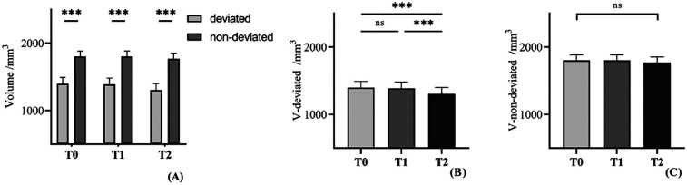
Results: The condyle on the deviated side was smaller than that on the non-deviated side, with significant volume reductions observed six months post-surgery on the deviated side. Several condylar changes were observed immediately after surgery, though of small magnitude, and it mostly tended to return to their original positions 6 months after surgery. However, the condyle rotated laterally on the deviated side and medially on the non-deviated side post operation and in long-term.
Conclusion: For patients with asymmetry, condyle on the deviated side undergo greater remodeling than the non-deviated side after orthognathic surgery. There are measurable rotations in the coronal plane of condyle on both sides.
期刊介绍:
Evidence of surgical interventions go back to prehistoric times. Since then, the field of surgery has developed into a complex array of specialties and procedures, particularly with the advent of microsurgery, lasers and minimally invasive techniques. The advanced skills now required from surgeons has led to ever increasing specialization, though these still share important fundamental principles.
Frontiers in Surgery is the umbrella journal representing the publication interests of all surgical specialties. It is divided into several “Specialty Sections” listed below. All these sections have their own Specialty Chief Editor, Editorial Board and homepage, but all articles carry the citation Frontiers in Surgery.
Frontiers in Surgery calls upon medical professionals and scientists from all surgical specialties to publish their experimental and clinical studies in this journal. By assembling all surgical specialties, which nonetheless retain their independence, under the common umbrella of Frontiers in Surgery, a powerful publication venue is created. Since there is often overlap and common ground between the different surgical specialties, assembly of all surgical disciplines into a single journal will foster a collaborative dialogue amongst the surgical community. This means that publications, which are also of interest to other surgical specialties, will reach a wider audience and have greater impact.
The aim of this multidisciplinary journal is to create a discussion and knowledge platform of advances and research findings in surgical practice today to continuously improve clinical management of patients and foster innovation in this field.

 求助内容:
求助内容: 应助结果提醒方式:


