Synthesis and characterization of novel silver vanadate nanofibers and consideration of the effects on MCF-7 and HDF cell line
IF 5.4
3区 化学
Q1 CHEMISTRY, INORGANIC & NUCLEAR
引用次数: 0
Abstract
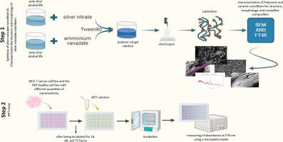
With the aim of introducing novel anticancer ceramic nanofibers, silver vanadate (AgVO3) nanofibers were successfully created by electrospinning and calcination in the search for novel anticancer ceramic nanofibers. Originally, a homogenous solution including silver nitrate, ammonium metavanadate, and poly vinyl alcohol was electrosppined to create composite polymer nanofibers. Silver vanadate ceramic nanofibers were created by calcining these composite nanofibers thereafter. Field emission-scanning electron microscopy (FE-SEM), energy dispersive X-ray spectroscopy (EDAX), elemental mapping analysis (EMPA), Fourier transform infrared spectroscopy (FT-IR), and X-ray powder diffraction (XRD) were used to characterize the produced nanofibers.
The analysis verified that composite nanofibers with a smooth surface and homogeneous shape had been successfully electrosppined. The precursor was annealed at 300 °C for three hours, resulting in well-crystallized fiber-like morphology in the ceramic nanostructures, which had diameters ranging from 40 to 120 nm. By using elemental mapping analysis and EDAX, the presence of V, O, and Ag elements was confirmed. Diffraction peaks consistent with silver vanadate's monoclinic crystal structure were found by XRD investigation.
The MTT assay results showed that silver vanadate nanofibers exhibited a time-dependent cytotoxic effect on MCF-7 cells with IC50 values of 28.2 μM (24 h), 10.82 μM (48 h), and 4.906 μM (72 h). In contrast, higher IC50 values were observed in HDF cells, including 569.5 μM (24 h), 181.5 μM (48 h), and 153.2 μM (72 h), indicating a favorable selectivity toward cancer cells. These findings offer new insights into the potential application of ceramic nanofibers for targeted cancer therapy.
新型钒酸银纳米纤维的合成、表征及对MCF-7和HDF细胞系的影响
以研制新型抗癌陶瓷纳米纤维为目的,通过静电纺丝和煅烧制备钒酸银纳米纤维,探索新型抗癌陶瓷纳米纤维。最初,一种含有硝酸银、偏氰酸铵和聚乙烯醇的均质溶液被电纺丝制成复合聚合物纳米纤维。然后将这些复合纳米纤维煅烧制成钒酸银陶瓷纳米纤维。利用场发射扫描电镜(FE-SEM)、能量色散x射线能谱(EDAX)、元素映射分析(EMPA)、傅里叶变换红外光谱(FT-IR)和x射线粉末衍射(XRD)对制备的纳米纤维进行了表征。结果表明,电纺丝成功制备出表面光滑、形状均匀的复合纳米纤维。前驱体在300℃下退火3小时,在直径为40 ~ 120 nm的陶瓷纳米结构中形成了结晶良好的纤维状结构。通过元素映射分析和EDAX分析,确定了样品中存在V、O和Ag元素。XRD分析发现了符合钒酸银单斜晶结构的衍射峰。MTT实验结果表明,钒酸银纳米纤维对MCF-7细胞的IC50值分别为28.2 μM (24 h)、10.82 μM (48 h)和4.906 μM (72 h),具有时间依赖性。相比之下,HDF细胞的IC50值更高,分别为569.5 μM (24 h)、181.5 μM (48 h)和153.2 μM (72 h),表明其对癌细胞具有良好的选择性。这些发现为陶瓷纳米纤维在靶向癌症治疗中的潜在应用提供了新的见解。
本文章由计算机程序翻译,如有差异,请以英文原文为准。
求助全文
约1分钟内获得全文
求助全文
来源期刊
Inorganic Chemistry Communications
化学-无机化学与核化学
CiteScore
5.50
自引率
7.90%
发文量
1013
审稿时长
53 days
期刊介绍:
Launched in January 1998, Inorganic Chemistry Communications is an international journal dedicated to the rapid publication of short communications in the major areas of inorganic, organometallic and supramolecular chemistry. Topics include synthetic and reaction chemistry, kinetics and mechanisms of reactions, bioinorganic chemistry, photochemistry and the use of metal and organometallic compounds in stoichiometric and catalytic synthesis or organic compounds.
 求助内容:
求助内容: 应助结果提醒方式:


