R Gervasi, G L Piazzetta, G Soluri, C Scigliano, C Pelaia, N Lobello, E Allegra, E Chiarella, N Innaro
{"title":"A rare case of supernumerary and ectopic parathyroid adenoma in the parotid gland: diagnostic and surgical challenges.","authors":"R Gervasi, G L Piazzetta, G Soluri, C Scigliano, C Pelaia, N Lobello, E Allegra, E Chiarella, N Innaro","doi":"10.1186/s13000-025-01712-4","DOIUrl":null,"url":null,"abstract":"<p><strong>Introduction: </strong>Primary hyperparathyroidism (PHPT) is a prevalent endocrine disorder characterized by elevated parathyroid hormone (PTH) levels and hypercalcemia, most commonly caused by solitary adenomas. Double adenomas, particularly those arising in ectopic and supernumerary glands, represent a rare diagnostic and surgical challenge.</p><p><strong>Case presentation: </strong>We report the case of a 64-year-old woman presenting with symptomatic PHPT. Preoperative imaging demonstrated uptake consistent with two hyperfunctioning parathyroid adenomas, including a rare supernumerary ectopic adenoma in lesion the right parotid region. Definitive diagnosis and surgical planning were guided by 18 F-fluorocholine PET/CT, which proved superior to conventional modalities.</p><p><strong>Discussion: </strong>This case underscores the critical role of advanced imaging techniques in the localization of parathyroid adenomas, particularly in anatomically atypical sites. The combination of functional and anatomical imaging with 18 F-fluorocholine PET/CT enabled accurate detection of both lesions and informed a multidisciplinary surgical approach.</p><p><strong>Conclusion: </strong>Integration of 18 F-fluorocholine PET/CT into the diagnostic workflow enhances the precision of parathyroid adenoma localization, especially in rare ectopic presentations. This contributes to tailored surgical strategies and improved patient outcomes. Histopathological examination confirmed two distinct adenomas, including one embedded in the parotid gland, supporting the diagnosis of a supernumerary ectopic parathyroid adenoma.</p>","PeriodicalId":11237,"journal":{"name":"Diagnostic Pathology","volume":"20 1","pages":"110"},"PeriodicalIF":2.3000,"publicationDate":"2025-10-02","publicationTypes":"Journal Article","fieldsOfStudy":null,"isOpenAccess":false,"openAccessPdf":"https://www.ncbi.nlm.nih.gov/pmc/articles/PMC12492863/pdf/","citationCount":"0","resultStr":null,"platform":"Semanticscholar","paperid":null,"PeriodicalName":"Diagnostic Pathology","FirstCategoryId":"3","ListUrlMain":"https://doi.org/10.1186/s13000-025-01712-4","RegionNum":3,"RegionCategory":"医学","ArticlePicture":[],"TitleCN":null,"AbstractTextCN":null,"PMCID":null,"EPubDate":"","PubModel":"","JCR":"Q2","JCRName":"PATHOLOGY","Score":null,"Total":0}
引用次数: 0
Abstract
Introduction: Primary hyperparathyroidism (PHPT) is a prevalent endocrine disorder characterized by elevated parathyroid hormone (PTH) levels and hypercalcemia, most commonly caused by solitary adenomas. Double adenomas, particularly those arising in ectopic and supernumerary glands, represent a rare diagnostic and surgical challenge.
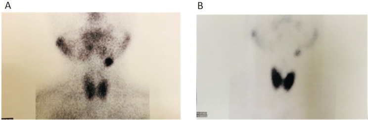
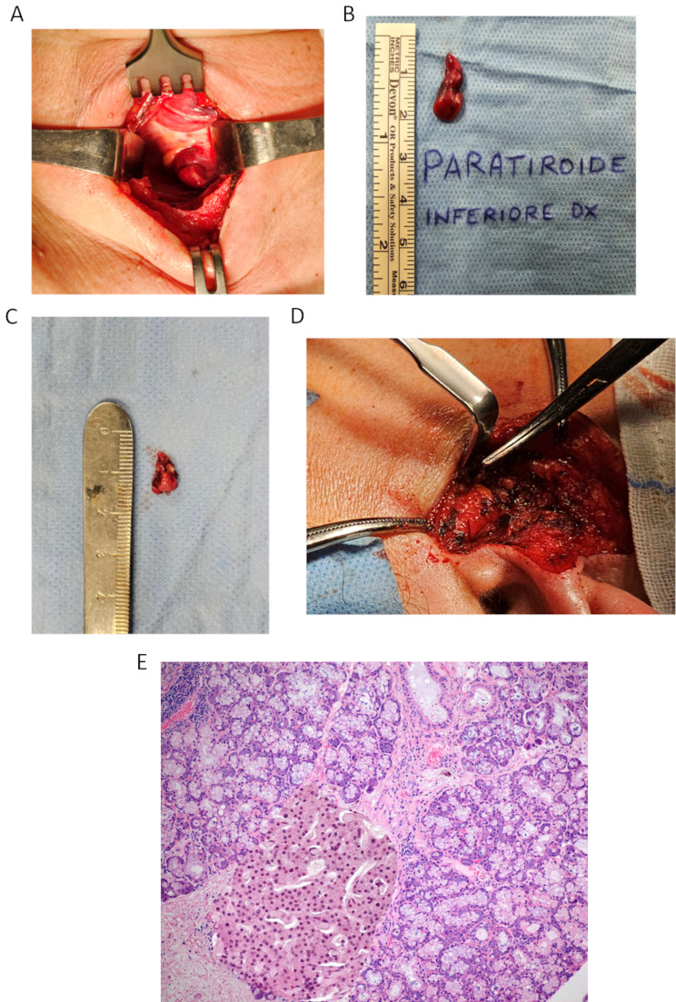
Case presentation: We report the case of a 64-year-old woman presenting with symptomatic PHPT. Preoperative imaging demonstrated uptake consistent with two hyperfunctioning parathyroid adenomas, including a rare supernumerary ectopic adenoma in lesion the right parotid region. Definitive diagnosis and surgical planning were guided by 18 F-fluorocholine PET/CT, which proved superior to conventional modalities.
Discussion: This case underscores the critical role of advanced imaging techniques in the localization of parathyroid adenomas, particularly in anatomically atypical sites. The combination of functional and anatomical imaging with 18 F-fluorocholine PET/CT enabled accurate detection of both lesions and informed a multidisciplinary surgical approach.
Conclusion: Integration of 18 F-fluorocholine PET/CT into the diagnostic workflow enhances the precision of parathyroid adenoma localization, especially in rare ectopic presentations. This contributes to tailored surgical strategies and improved patient outcomes. Histopathological examination confirmed two distinct adenomas, including one embedded in the parotid gland, supporting the diagnosis of a supernumerary ectopic parathyroid adenoma.
原发性甲状旁腺功能亢进(PHPT)是一种常见的内分泌疾病,以甲状旁腺激素(PTH)水平升高和高钙血症为特征,最常由孤立腺瘤引起。双腺瘤,特别是那些发生在异位腺和多余腺,是一种罕见的诊断和手术挑战。病例介绍:我们报告的情况下,64岁的妇女提出症状PHPT。术前影像学显示摄取与两个功能亢进的甲状旁腺瘤一致,包括一个罕见的右侧腮腺区病变的外生异位腺瘤。最终诊断和手术计划由18f -氟胆碱PET/CT指导,证明其优于传统方式。讨论:本病例强调了先进成像技术在甲状旁腺瘤定位中的关键作用,特别是在解剖上不典型的部位。18 f -氟胆碱PET/CT结合功能和解剖成像,能够准确检测这两种病变,并为多学科手术方法提供信息。结论:将18f -氟胆碱PET/CT整合到诊断流程中,可提高甲状旁腺瘤定位的准确性,特别是在罕见的异位表现中。这有助于定制手术策略并改善患者预后。组织病理学检查证实两种不同的腺瘤,包括一种嵌入腮腺,支持多余异位甲状旁腺瘤的诊断。
期刊介绍:
Diagnostic Pathology is an open access, peer-reviewed, online journal that considers research in surgical and clinical pathology, immunology, and biology, with a special focus on cutting-edge approaches in diagnostic pathology and tissue-based therapy. The journal covers all aspects of surgical pathology, including classic diagnostic pathology, prognosis-related diagnosis (tumor stages, prognosis markers, such as MIB-percentage, hormone receptors, etc.), and therapy-related findings. The journal also focuses on the technological aspects of pathology, including molecular biology techniques, morphometry aspects (stereology, DNA analysis, syntactic structure analysis), communication aspects (telecommunication, virtual microscopy, virtual pathology institutions, etc.), and electronic education and quality assurance (for example interactive publication, on-line references with automated updating, etc.).

 求助内容:
求助内容: 应助结果提醒方式:


