Rohit Pardasani, Renee Vitullo, Sara Harris, Halit O Yapici, John Beard
{"title":"Development of a novel artificial intelligence algorithm for interpreting fetal heart rate and uterine activity data in cardiotocography.","authors":"Rohit Pardasani, Renee Vitullo, Sara Harris, Halit O Yapici, John Beard","doi":"10.3389/fdgth.2025.1638424","DOIUrl":null,"url":null,"abstract":"<p><strong>Introduction: </strong>Cardiotocography (CTG) assesses fetal well-being through measurements of fetal heart rate (FHR) and uterine activity (UA). Manual visual assessment of fetal tracings is variable due to the subjective nature of their interpretation. Artificial intelligence (AI) using automatic signal processing may be leveraged to support consistent, comprehensive interpretations. This study demonstrated the development and training of a novel AI algorithm that analyzes and interprets certain clinical events and parameters calculated during labor to assist with clinical decisions.</p><p><strong>Methods: </strong>Fetal tracings sourced from 19 birthing centers through a US-based healthcare delivery organization were clinically interpreted, labeled, quality checked, and ratified by clinicians to be included in the study. The algorithm using deep learning and rule-based techniques was developed to identify segments of interest (accelerations, decelerations, and contractions). A three parallel one-dimensional Unet design with two inputs (FHR and UA) and one channel output each (for accelerations, decelerations, and contractions) was selected as the final architecture. Algorithm performance was evaluated through recall (sensitivity), precision, <i>F</i>1 score, and duration and numerical ratios.</p><p><strong>Results: </strong>A total of 133,696 patient files were used to create fetal tracings. After the exclusion, labeling, and ratification processes, the final datasets included 1,600 tracings for training, 421 for validation, and 591 for testing. The model provided promising performance and achieved <i>F</i>1 scores of 0.803 for accelerations, 0.520 for decelerations, and 0.868 for contractions on the final test set, with a 91.5% predicted baseline accuracy (difference of ≤5 bpm) compared to clinician interpretation.</p><p><strong>Conclusion: </strong>This study demonstrates the successful development of a novel AI algorithm utilizing FHR and UA data to analyze and interpret fetal tracing events and parameters. The algorithm may have potential to enhance patient care by supporting bedside clinician CTG interpretation.</p>","PeriodicalId":73078,"journal":{"name":"Frontiers in digital health","volume":"7 ","pages":"1638424"},"PeriodicalIF":3.2000,"publicationDate":"2025-09-16","publicationTypes":"Journal Article","fieldsOfStudy":null,"isOpenAccess":false,"openAccessPdf":"https://www.ncbi.nlm.nih.gov/pmc/articles/PMC12479546/pdf/","citationCount":"0","resultStr":null,"platform":"Semanticscholar","paperid":null,"PeriodicalName":"Frontiers in digital health","FirstCategoryId":"1085","ListUrlMain":"https://doi.org/10.3389/fdgth.2025.1638424","RegionNum":0,"RegionCategory":null,"ArticlePicture":[],"TitleCN":null,"AbstractTextCN":null,"PMCID":null,"EPubDate":"2025/1/1 0:00:00","PubModel":"eCollection","JCR":"Q1","JCRName":"HEALTH CARE SCIENCES & SERVICES","Score":null,"Total":0}
引用次数: 0
Abstract
Introduction: Cardiotocography (CTG) assesses fetal well-being through measurements of fetal heart rate (FHR) and uterine activity (UA). Manual visual assessment of fetal tracings is variable due to the subjective nature of their interpretation. Artificial intelligence (AI) using automatic signal processing may be leveraged to support consistent, comprehensive interpretations. This study demonstrated the development and training of a novel AI algorithm that analyzes and interprets certain clinical events and parameters calculated during labor to assist with clinical decisions.
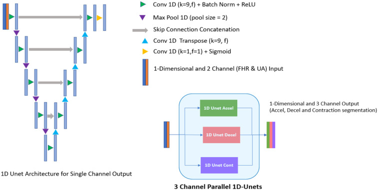
Methods: Fetal tracings sourced from 19 birthing centers through a US-based healthcare delivery organization were clinically interpreted, labeled, quality checked, and ratified by clinicians to be included in the study. The algorithm using deep learning and rule-based techniques was developed to identify segments of interest (accelerations, decelerations, and contractions). A three parallel one-dimensional Unet design with two inputs (FHR and UA) and one channel output each (for accelerations, decelerations, and contractions) was selected as the final architecture. Algorithm performance was evaluated through recall (sensitivity), precision, F1 score, and duration and numerical ratios.
Results: A total of 133,696 patient files were used to create fetal tracings. After the exclusion, labeling, and ratification processes, the final datasets included 1,600 tracings for training, 421 for validation, and 591 for testing. The model provided promising performance and achieved F1 scores of 0.803 for accelerations, 0.520 for decelerations, and 0.868 for contractions on the final test set, with a 91.5% predicted baseline accuracy (difference of ≤5 bpm) compared to clinician interpretation.
Conclusion: This study demonstrates the successful development of a novel AI algorithm utilizing FHR and UA data to analyze and interpret fetal tracing events and parameters. The algorithm may have potential to enhance patient care by supporting bedside clinician CTG interpretation.

 求助内容:
求助内容: 应助结果提醒方式:


