{"title":"Changes in Visual Evoked Potential and Optical Coherence Tomography in Parkinson's Disease: A Systematic Review and Meta-Analysis.","authors":"Zahra Hemmatian, Javad Heravian Shandiz, Ali Shoeibi, Nasser Shoeibi, Reyhane Shariati, Batool Haghighi, Firozeh Fereydouni, Negareh Yazdani","doi":"10.1155/padi/2386302","DOIUrl":null,"url":null,"abstract":"<p><p><b>Background:</b> Previous studies revealed that optical coherence tomography (OCT) and visual evoked potential (VEP) were impaired in patients with Parkinson's disease (PD), but the results were inconsistent; in this meta-analysis, we tried to answer this issue by including studies that performed these two tests on the same sample size. <b>Methods:</b> PubMed, Scopus, Cochrane, and Google Scholar were comprehensively reviewed to retrieve the published studies investigating changes in OCT and VEP responses in PD patients. We analyzed the pooled weighted difference in means between PD patients and healthy controls using the random-effects model. <b>Results:</b> Ten studies were included (12 sets of data), enrolling 337 PD patients and 273 healthy controls. The P100 latency in PD patients was significantly higher compared to healthy controls (difference in means = 6.16, 95% CI: 1.16-11.15, <i>p</i>=0.02, <i>n</i> = 11). Significant thinning of the retinal nerve fiber layer (difference in means = -4.38, 95% CI: -6.29 to -2.47, <i>p</i> ≤ 0.001, <i>n</i> = 11) was observed in the PD eyes compared to the healthy subjects. However, no statistically significant difference was found in the means of P100 amplitude (<i>p</i>=0.06) and the average central foveal thickness (<i>p</i>=0.08) between PD patients and the control group. There was a significant negative correlation between RNFL weighted mean difference and P100 latency (<i>r</i> = -0.65, <i>p</i> ≤ 0.001) in all subjects. <b>Conclusions:</b> Our results confirmed that Parkinson's patients showed significant thinning of RNFL thickness and prolonged P100 latency time.</p>","PeriodicalId":19907,"journal":{"name":"Parkinson's Disease","volume":"2025 ","pages":"2386302"},"PeriodicalIF":2.2000,"publicationDate":"2025-09-23","publicationTypes":"Journal Article","fieldsOfStudy":null,"isOpenAccess":false,"openAccessPdf":"https://www.ncbi.nlm.nih.gov/pmc/articles/PMC12483749/pdf/","citationCount":"0","resultStr":null,"platform":"Semanticscholar","paperid":null,"PeriodicalName":"Parkinson's Disease","FirstCategoryId":"3","ListUrlMain":"https://doi.org/10.1155/padi/2386302","RegionNum":4,"RegionCategory":"医学","ArticlePicture":[],"TitleCN":null,"AbstractTextCN":null,"PMCID":null,"EPubDate":"2025/1/1 0:00:00","PubModel":"eCollection","JCR":"Q3","JCRName":"CLINICAL NEUROLOGY","Score":null,"Total":0}
引用次数: 0
Abstract
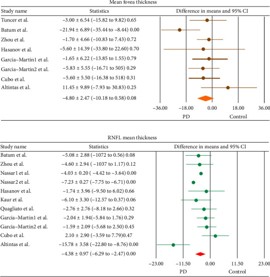
Background: Previous studies revealed that optical coherence tomography (OCT) and visual evoked potential (VEP) were impaired in patients with Parkinson's disease (PD), but the results were inconsistent; in this meta-analysis, we tried to answer this issue by including studies that performed these two tests on the same sample size. Methods: PubMed, Scopus, Cochrane, and Google Scholar were comprehensively reviewed to retrieve the published studies investigating changes in OCT and VEP responses in PD patients. We analyzed the pooled weighted difference in means between PD patients and healthy controls using the random-effects model. Results: Ten studies were included (12 sets of data), enrolling 337 PD patients and 273 healthy controls. The P100 latency in PD patients was significantly higher compared to healthy controls (difference in means = 6.16, 95% CI: 1.16-11.15, p=0.02, n = 11). Significant thinning of the retinal nerve fiber layer (difference in means = -4.38, 95% CI: -6.29 to -2.47, p ≤ 0.001, n = 11) was observed in the PD eyes compared to the healthy subjects. However, no statistically significant difference was found in the means of P100 amplitude (p=0.06) and the average central foveal thickness (p=0.08) between PD patients and the control group. There was a significant negative correlation between RNFL weighted mean difference and P100 latency (r = -0.65, p ≤ 0.001) in all subjects. Conclusions: Our results confirmed that Parkinson's patients showed significant thinning of RNFL thickness and prolonged P100 latency time.
期刊介绍:
Parkinson’s Disease is a peer-reviewed, Open Access journal that publishes original research articles, review articles, and clinical studies related to the epidemiology, etiology, pathogenesis, genetics, cellular, molecular and neurophysiology, as well as the diagnosis and treatment of Parkinson’s disease.


 求助内容:
求助内容: 应助结果提醒方式:


