Bogusz Aksak-Wąs, Karolina Skonieczna-Żydecka, Miłosz Parczewski, Rafał Hrynkiewicz, Filip Lewandowski, Karol Serwin, Kaja Mielczak, Adam Majchrzak, Franciszek Lenkiewicz, Paulina Niedźwiedzka-Rystwej, Poorani Gurumallesh
{"title":"Influence of PD-1 and PD-1L Immune Exhaustion Receptors on Immune Reconstruction in People Living With HIV.","authors":"Bogusz Aksak-Wąs, Karolina Skonieczna-Żydecka, Miłosz Parczewski, Rafał Hrynkiewicz, Filip Lewandowski, Karol Serwin, Kaja Mielczak, Adam Majchrzak, Franciszek Lenkiewicz, Paulina Niedźwiedzka-Rystwej, Poorani Gurumallesh","doi":"10.1155/jimr/2462382","DOIUrl":null,"url":null,"abstract":"<p><strong>Introduction: </strong>The progressive immunological impairment associated with human immunodeficiency virus (HIV) infection is partially mediated by the programmed cell death protein-1 (PD-1)/programed death-ligand 1(PD-L1) inhibitory pathway. This investigation aims to evaluate the influence of PD-1 on immune reconstitution in patients undergoing antiretroviral therapy (ART), with data visualized through principal component analysis (PCA).</p><p><strong>Materials and methods: </strong>Data from 52 ART-treated individuals achieving viral suppression were analyzed over 12 months. CD4+, CD8+, CD19+, and PD-1/PD-L1 expressions were quantified via flow cytometry at baseline and after 12 months, and immune recovery was assessed at CD4+ thresholds of 500 and 800/μL and CD4+/CD8+ ratios of >0.8 and >1.0 using linear and logistic regression. PCA was applied to visualize clustering of immune recovery patterns based on PD-1/PD-L1 expression levels and immune cell counts, with statistical significance evaluated using ANOVA.</p><p><strong>Results: </strong>The analyzed group of 52 patients was predominantly male (65.4%; <i>n</i> = 34). PD-1/PD-L1 expression showed modest associations with immune recovery. Higher PD-L1 expression on CD3+ T-cells at baseline was associated with a reduced likelihood of recovery to CD4+>500/μL (OR: 0.79; 95%CI: 0.62-0.99; <i>p</i> = 0.04). Linear regression demonstrated that increased PD-L1 on CD4+ T-cells and PD-1 on CD19+ B-cells positively correlated with higher CD4+/CD8+ ratios at follow-up (coefficient: 0.035 and 0.03, respectively; <i>p</i> < 0.02), while logistic regression indicated that higher PD-1 on CD3+ T-cells increased the odds of recovery to CD4+>500/μL (OR: 1.03; 95% CI: 1.0036-1.07); = 0.03). Notably, this weak signal may result from a general increase in the number of lymphocytes during therapy. PCA did not reveal significant clustering of immune recovery patterns.</p><p><strong>Conclusion: </strong>PD-1 and PD-L1 expressions on immune cells are weakly associated with immune recovery metrics in individuals undergoing ART. Further research is needed to explore their role in immune reconstitution and potential clinical applications.</p>","PeriodicalId":15952,"journal":{"name":"Journal of Immunology Research","volume":"2025 ","pages":"2462382"},"PeriodicalIF":3.6000,"publicationDate":"2025-09-30","publicationTypes":"Journal Article","fieldsOfStudy":null,"isOpenAccess":false,"openAccessPdf":"https://www.ncbi.nlm.nih.gov/pmc/articles/PMC12481824/pdf/","citationCount":"0","resultStr":null,"platform":"Semanticscholar","paperid":null,"PeriodicalName":"Journal of Immunology Research","FirstCategoryId":"3","ListUrlMain":"https://doi.org/10.1155/jimr/2462382","RegionNum":3,"RegionCategory":"医学","ArticlePicture":[],"TitleCN":null,"AbstractTextCN":null,"PMCID":null,"EPubDate":"2025/1/1 0:00:00","PubModel":"eCollection","JCR":"Q2","JCRName":"IMMUNOLOGY","Score":null,"Total":0}
引用次数: 0
Abstract
Introduction: The progressive immunological impairment associated with human immunodeficiency virus (HIV) infection is partially mediated by the programmed cell death protein-1 (PD-1)/programed death-ligand 1(PD-L1) inhibitory pathway. This investigation aims to evaluate the influence of PD-1 on immune reconstitution in patients undergoing antiretroviral therapy (ART), with data visualized through principal component analysis (PCA).
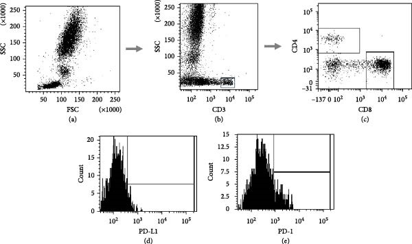
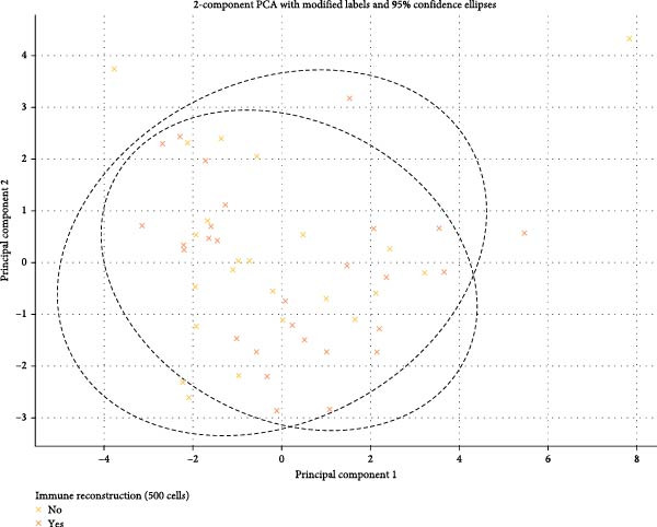
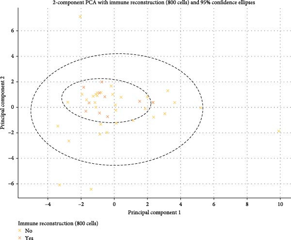
Materials and methods: Data from 52 ART-treated individuals achieving viral suppression were analyzed over 12 months. CD4+, CD8+, CD19+, and PD-1/PD-L1 expressions were quantified via flow cytometry at baseline and after 12 months, and immune recovery was assessed at CD4+ thresholds of 500 and 800/μL and CD4+/CD8+ ratios of >0.8 and >1.0 using linear and logistic regression. PCA was applied to visualize clustering of immune recovery patterns based on PD-1/PD-L1 expression levels and immune cell counts, with statistical significance evaluated using ANOVA.
Results: The analyzed group of 52 patients was predominantly male (65.4%; n = 34). PD-1/PD-L1 expression showed modest associations with immune recovery. Higher PD-L1 expression on CD3+ T-cells at baseline was associated with a reduced likelihood of recovery to CD4+>500/μL (OR: 0.79; 95%CI: 0.62-0.99; p = 0.04). Linear regression demonstrated that increased PD-L1 on CD4+ T-cells and PD-1 on CD19+ B-cells positively correlated with higher CD4+/CD8+ ratios at follow-up (coefficient: 0.035 and 0.03, respectively; p < 0.02), while logistic regression indicated that higher PD-1 on CD3+ T-cells increased the odds of recovery to CD4+>500/μL (OR: 1.03; 95% CI: 1.0036-1.07); = 0.03). Notably, this weak signal may result from a general increase in the number of lymphocytes during therapy. PCA did not reveal significant clustering of immune recovery patterns.
Conclusion: PD-1 and PD-L1 expressions on immune cells are weakly associated with immune recovery metrics in individuals undergoing ART. Further research is needed to explore their role in immune reconstitution and potential clinical applications.
期刊介绍:
Journal of Immunology Research is a peer-reviewed, Open Access journal that provides a platform for scientists and clinicians working in different areas of immunology and therapy. The journal publishes research articles, review articles, as well as clinical studies related to classical immunology, molecular immunology, clinical immunology, cancer immunology, transplantation immunology, immune pathology, immunodeficiency, autoimmune diseases, immune disorders, and immunotherapy.
 求助内容:
求助内容: 应助结果提醒方式:


