Recurrent 0 μm vault complicated with anterior subcapsular cataract after toric ICL in an eye with multiple anatomical risk factors: clinical reflections.
{"title":"Recurrent 0 μm vault complicated with anterior subcapsular cataract after toric ICL in an eye with multiple anatomical risk factors: clinical reflections.","authors":"Yue-Xin Chen, Xue-Yan Li, Yan-Ying Zhu, Yu-Kun Liu, Hai-Yan Xie, Xiao-Chen Xu, Jing Wang","doi":"10.1186/s12886-025-04343-x","DOIUrl":null,"url":null,"abstract":"<p><strong>Background: </strong>This is a case of a patient who developed 0 μm vault after TICL implantation with concomitant anterior subcapsular cataract, and 0 μm vault again after TICL replacement.</p><p><strong>Case presentation: </strong>A 19-year-old woman with high myopia underwent bilateral TICL implantation. Preoperative imaging revealed shallow anterior chambers, high crystalline lens rise, small pupils in Pentacam HR (Oculus, Germany), and short ciliary processes in ultrasound biomicroscopy (UBM). Both eyes developed low postoperative vaults. Nineteen months later, she returned with bilateral 0 μm vault and anterior subcapsular cataracts. Repeat UBM confirmed short ciliary processes and partial footplate slippage beneath the ciliary sulcus. The left TICL was replaced with a larger lens, but 0 μm vault recurred. The right eye was monitored without further intervention.</p><p><strong>Conclusions: </strong>Although factors like shallow anterior chamber, high lens rise, and small pupils can often be managed with proper lens sizing, this case suggests that certain anatomical features-such as abnormal ciliary body structure-may lead to persistently low or zero vault despite optimized planning. UBM is a valuable tool for identifying such hidden risks and should be included in preoperative assessment for high-risk patients.</p>","PeriodicalId":9058,"journal":{"name":"BMC Ophthalmology","volume":"25 1","pages":"528"},"PeriodicalIF":1.7000,"publicationDate":"2025-10-01","publicationTypes":"Journal Article","fieldsOfStudy":null,"isOpenAccess":false,"openAccessPdf":"https://www.ncbi.nlm.nih.gov/pmc/articles/PMC12486667/pdf/","citationCount":"0","resultStr":null,"platform":"Semanticscholar","paperid":null,"PeriodicalName":"BMC Ophthalmology","FirstCategoryId":"3","ListUrlMain":"https://doi.org/10.1186/s12886-025-04343-x","RegionNum":4,"RegionCategory":"医学","ArticlePicture":[],"TitleCN":null,"AbstractTextCN":null,"PMCID":null,"EPubDate":"","PubModel":"","JCR":"Q3","JCRName":"OPHTHALMOLOGY","Score":null,"Total":0}
引用次数: 0
Abstract
Background: This is a case of a patient who developed 0 μm vault after TICL implantation with concomitant anterior subcapsular cataract, and 0 μm vault again after TICL replacement.
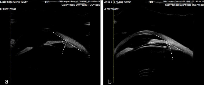
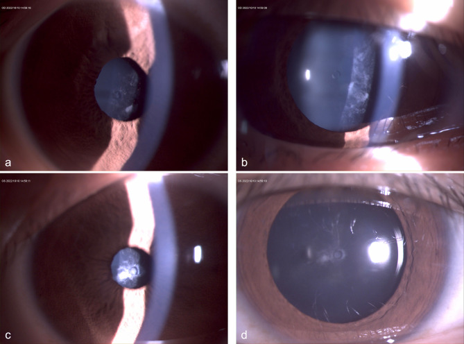
Case presentation: A 19-year-old woman with high myopia underwent bilateral TICL implantation. Preoperative imaging revealed shallow anterior chambers, high crystalline lens rise, small pupils in Pentacam HR (Oculus, Germany), and short ciliary processes in ultrasound biomicroscopy (UBM). Both eyes developed low postoperative vaults. Nineteen months later, she returned with bilateral 0 μm vault and anterior subcapsular cataracts. Repeat UBM confirmed short ciliary processes and partial footplate slippage beneath the ciliary sulcus. The left TICL was replaced with a larger lens, but 0 μm vault recurred. The right eye was monitored without further intervention.
Conclusions: Although factors like shallow anterior chamber, high lens rise, and small pupils can often be managed with proper lens sizing, this case suggests that certain anatomical features-such as abnormal ciliary body structure-may lead to persistently low or zero vault despite optimized planning. UBM is a valuable tool for identifying such hidden risks and should be included in preoperative assessment for high-risk patients.
期刊介绍:
BMC Ophthalmology is an open access, peer-reviewed journal that considers articles on all aspects of the prevention, diagnosis and management of eye disorders, as well as related molecular genetics, pathophysiology, and epidemiology.

 求助内容:
求助内容: 应助结果提醒方式:


