Baqir J Kedwai, Zachary R Zottola, Daniel J Lehane, Joshua T Geiger, Micheal C Stoner, Michael S Richards, Doran S Mix
{"title":"Characterizing changes in abdominal aortic aneurysms using principal wall strain ultrasound elastography.","authors":"Baqir J Kedwai, Zachary R Zottola, Daniel J Lehane, Joshua T Geiger, Micheal C Stoner, Michael S Richards, Doran S Mix","doi":"10.3389/fcvm.2025.1613881","DOIUrl":null,"url":null,"abstract":"<p><strong>Introduction: </strong>Aortic principal wall strain is a biomechanical parameter correlated with aneurysm growth rate that affects abdominal aortic aneurysm (AAA) stability. Characterize changes in pressure-normalized maximum mean aortic principal wall strain <math><mo>(</mo> <mrow> <mover><msub><mi>ε</mi> <mrow><mi>ρ</mi> <mo>+</mo></mrow> </msub> <mo>¯</mo></mover> <mrow><mo>/</mo> <mi>PP</mi></mrow> </mrow> <mo>)</mo></math> using ultrasound elastography (USE).</p><p><strong>Methods: </strong>Axial ultrasound images of patient AAAs were collected at two consecutive clinic visits. The <math> <mover><msub><mi>ε</mi> <mrow><mi>ρ</mi> <mo>+</mo></mrow> </msub> <mo>¯</mo></mover> <mrow><mo>/</mo> <mi>PP</mi></mrow> </math> for each image was calculated using a novel finite element mesh technique. The cohort was separated by index <math> <mover><msub><mi>ε</mi> <mrow><mi>ρ</mi> <mo>+</mo></mrow> </msub> <mo>¯</mo></mover> <mrow><mo>/</mo> <mi>PP</mi></mrow> </math> terciles, and the rate of strain change, growth, intervention, and rupture were compared.</p><p><strong>Results: </strong>31 patients with a median age of 72.0 [65.0, 77.5] at index visits were included, with follow-up imaging taken at an average interval of 6.2 [6.0, 8.3] months. For the whole cohort, maximum <math> <mover><msub><mi>ε</mi> <mrow><mi>ρ</mi> <mo>+</mo></mrow> </msub> <mo>¯</mo></mover> <mrow><mo>/</mo> <mi>PP</mi></mrow> </math> decreased from 2.1 [1.1, 2.7] %/mmHg to 1.9 [1.3, 2.6] %/mmHg (<i>p</i> = 0.08), and maximum AAA diameter increased from a median of 4.3 [4.0, 4.7] cm to 4.4 [4.1, 4.9] cm (<i>p</i> = 0.04). The \"high-strain\" tercile was associated with a strain reduction of -1.3 [-2.5, -1.1] %/mmHg between index and follow-up imaging, as compared to the \"low-strain\" (-0.1 [-0.6, 0.5] %/mmHg, <i>p</i> < 0.01) and \"intermediate-strain\" (-0.4 [-0.5, -0.3] %/mmHg, <i>p</i> = 0.04) terciles. There was no difference in the rate of AAA growth, intervention, or rupture between terciles.</p><p><strong>Discussion: </strong>The present findings indicate that <math> <mover><msub><mi>ε</mi> <mrow><mi>ρ</mi> <mo>+</mo></mrow> </msub> <mo>¯</mo></mover> <mrow><mo>/</mo> <mi>PP</mi></mrow> </math> at baseline predicts the degree and direction of <math> <mover><msub><mi>ε</mi> <mrow><mi>ρ</mi> <mo>+</mo></mrow> </msub> <mo>¯</mo></mover> <mrow><mo>/</mo> <mi>PP</mi></mrow> </math> change in AAAs over time. These findings offer insight into the natural history of AAA tissue mechanics and demonstrate the potential for a novel ultrasound technique to quantify biomechanical changes in the aortic wall. These findings may aid in the development of patient-specific risk stratification tools informed by biomechanical data in addition to conventional size-based criteria.</p>","PeriodicalId":12414,"journal":{"name":"Frontiers in Cardiovascular Medicine","volume":"12 ","pages":"1613881"},"PeriodicalIF":2.8000,"publicationDate":"2025-09-15","publicationTypes":"Journal Article","fieldsOfStudy":null,"isOpenAccess":false,"openAccessPdf":"https://www.ncbi.nlm.nih.gov/pmc/articles/PMC12477128/pdf/","citationCount":"0","resultStr":null,"platform":"Semanticscholar","paperid":null,"PeriodicalName":"Frontiers in Cardiovascular Medicine","FirstCategoryId":"3","ListUrlMain":"https://doi.org/10.3389/fcvm.2025.1613881","RegionNum":3,"RegionCategory":"医学","ArticlePicture":[],"TitleCN":null,"AbstractTextCN":null,"PMCID":null,"EPubDate":"2025/1/1 0:00:00","PubModel":"eCollection","JCR":"Q2","JCRName":"CARDIAC & CARDIOVASCULAR SYSTEMS","Score":null,"Total":0}
引用次数: 0
Abstract
Introduction: Aortic principal wall strain is a biomechanical parameter correlated with aneurysm growth rate that affects abdominal aortic aneurysm (AAA) stability. Characterize changes in pressure-normalized maximum mean aortic principal wall strain using ultrasound elastography (USE).
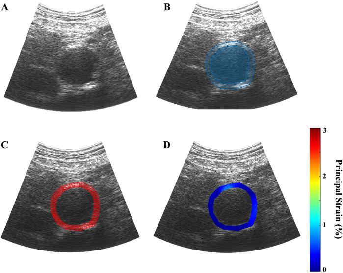
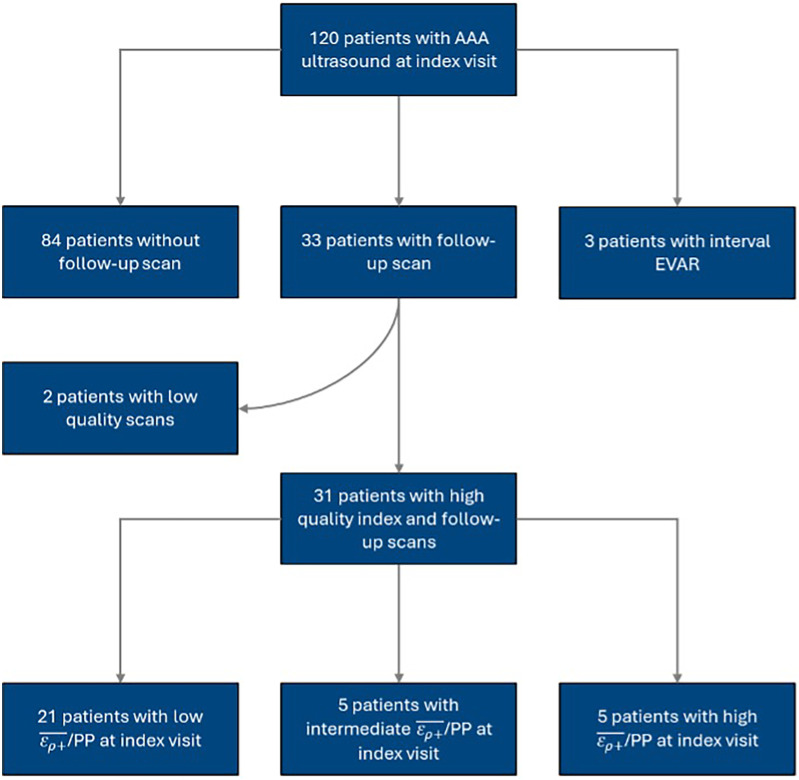
Methods: Axial ultrasound images of patient AAAs were collected at two consecutive clinic visits. The for each image was calculated using a novel finite element mesh technique. The cohort was separated by index terciles, and the rate of strain change, growth, intervention, and rupture were compared.
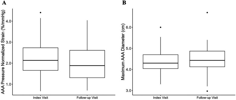
Results: 31 patients with a median age of 72.0 [65.0, 77.5] at index visits were included, with follow-up imaging taken at an average interval of 6.2 [6.0, 8.3] months. For the whole cohort, maximum decreased from 2.1 [1.1, 2.7] %/mmHg to 1.9 [1.3, 2.6] %/mmHg (p = 0.08), and maximum AAA diameter increased from a median of 4.3 [4.0, 4.7] cm to 4.4 [4.1, 4.9] cm (p = 0.04). The "high-strain" tercile was associated with a strain reduction of -1.3 [-2.5, -1.1] %/mmHg between index and follow-up imaging, as compared to the "low-strain" (-0.1 [-0.6, 0.5] %/mmHg, p < 0.01) and "intermediate-strain" (-0.4 [-0.5, -0.3] %/mmHg, p = 0.04) terciles. There was no difference in the rate of AAA growth, intervention, or rupture between terciles.
Discussion: The present findings indicate that at baseline predicts the degree and direction of change in AAAs over time. These findings offer insight into the natural history of AAA tissue mechanics and demonstrate the potential for a novel ultrasound technique to quantify biomechanical changes in the aortic wall. These findings may aid in the development of patient-specific risk stratification tools informed by biomechanical data in addition to conventional size-based criteria.
期刊介绍:
Frontiers? Which frontiers? Where exactly are the frontiers of cardiovascular medicine? And who should be defining these frontiers?
At Frontiers in Cardiovascular Medicine we believe it is worth being curious to foresee and explore beyond the current frontiers. In other words, we would like, through the articles published by our community journal Frontiers in Cardiovascular Medicine, to anticipate the future of cardiovascular medicine, and thus better prevent cardiovascular disorders and improve therapeutic options and outcomes of our patients.

 求助内容:
求助内容: 应助结果提醒方式:


