Mohamad Tlais, Hussein Hamze, Ali Hteit, Karim Haddad, Issam El Fassih, Issa Zalzali, Sally Mahmoud, Sabine Karaki, Diana Jabbour
{"title":"Advances in ultrasound-based imaging for diagnosis of endometrial cancer.","authors":"Mohamad Tlais, Hussein Hamze, Ali Hteit, Karim Haddad, Issam El Fassih, Issa Zalzali, Sally Mahmoud, Sabine Karaki, Diana Jabbour","doi":"10.4329/wjr.v17.i9.111493","DOIUrl":null,"url":null,"abstract":"<p><strong>Background: </strong>Endometrial cancer (EC) is the most common gynecological malignancy in high-income countries, with incidence rates rising globally. Early and accurate diagnosis is essential for improving outcomes. Transvaginal ultrasound (TVUS) remains a cost-effective first-line tool, and emerging techniques such as three-dimensional (3D) ultrasound (US), contrast-enhanced US (CEUS), elastography, and artificial intelligence (AI)-enhanced imaging may further improve diagnostic performance.</p><p><strong>Aim: </strong>To systematically review recent advances in US-based imaging techniques for the diagnosis and staging of EC, and to compare their performance with magnetic resonance imaging (MRI).</p><p><strong>Methods: </strong>A systematic search of PubMed, Scopus, Web of Science, and Google Scholar was performed to identify studies published between January 2010 and March 2025. Eligible studies evaluated TVUS, 3D-US, CEUS, elastography, or AI-enhanced US in EC diagnosis and staging. Methodological quality was assessed using the QUADAS-2 tool. Sensitivity, specificity, and area under the curve (AUC) were extracted where available, with narrative synthesis due to heterogeneity.</p><p><strong>Results: </strong>Forty-one studies met the inclusion criteria. TVUS demonstrated high sensitivity (76%-96%) but moderate specificity (61%-86%), while MRI achieved higher specificity (84%-95%) and superior staging accuracy. 3D-US yielded accuracy comparable to MRI in selected early-stage cases. CEUS and elastography enhanced tissue characterization, and AI-enhanced US achieved pooled AUCs up to 0.91 for risk prediction and lesion segmentation. Variability in performance was noted across modalities due to patient demographics, equipment differences, and operator experience.</p><p><strong>Conclusion: </strong>TVUS remains a highly sensitive initial screening tool, with MRI preferred for definitive staging. 3D-US, CEUS, elastography, and AI-enhanced techniques show promise as complementary or alternative approaches, particularly in low-resource settings. Standardization, multicenter validation, and integration of multi-modal imaging are needed to optimize diagnostic pathways for EC.</p>","PeriodicalId":23819,"journal":{"name":"World journal of radiology","volume":"17 9","pages":"111493"},"PeriodicalIF":1.5000,"publicationDate":"2025-09-28","publicationTypes":"Journal Article","fieldsOfStudy":null,"isOpenAccess":false,"openAccessPdf":"https://www.ncbi.nlm.nih.gov/pmc/articles/PMC12476809/pdf/","citationCount":"0","resultStr":null,"platform":"Semanticscholar","paperid":null,"PeriodicalName":"World journal of radiology","FirstCategoryId":"1085","ListUrlMain":"https://doi.org/10.4329/wjr.v17.i9.111493","RegionNum":0,"RegionCategory":null,"ArticlePicture":[],"TitleCN":null,"AbstractTextCN":null,"PMCID":null,"EPubDate":"","PubModel":"","JCR":"Q3","JCRName":"RADIOLOGY, NUCLEAR MEDICINE & MEDICAL IMAGING","Score":null,"Total":0}
引用次数: 0
Abstract
Background: Endometrial cancer (EC) is the most common gynecological malignancy in high-income countries, with incidence rates rising globally. Early and accurate diagnosis is essential for improving outcomes. Transvaginal ultrasound (TVUS) remains a cost-effective first-line tool, and emerging techniques such as three-dimensional (3D) ultrasound (US), contrast-enhanced US (CEUS), elastography, and artificial intelligence (AI)-enhanced imaging may further improve diagnostic performance.
Aim: To systematically review recent advances in US-based imaging techniques for the diagnosis and staging of EC, and to compare their performance with magnetic resonance imaging (MRI).
Methods: A systematic search of PubMed, Scopus, Web of Science, and Google Scholar was performed to identify studies published between January 2010 and March 2025. Eligible studies evaluated TVUS, 3D-US, CEUS, elastography, or AI-enhanced US in EC diagnosis and staging. Methodological quality was assessed using the QUADAS-2 tool. Sensitivity, specificity, and area under the curve (AUC) were extracted where available, with narrative synthesis due to heterogeneity.
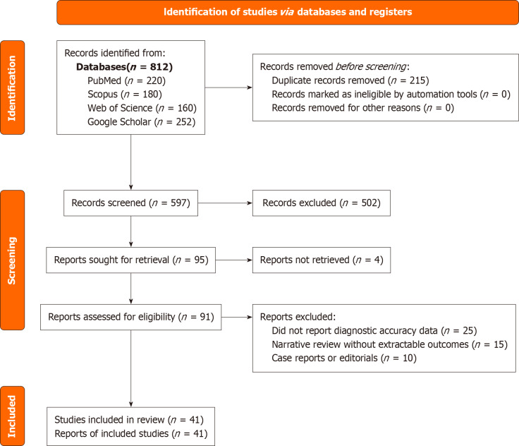
Results: Forty-one studies met the inclusion criteria. TVUS demonstrated high sensitivity (76%-96%) but moderate specificity (61%-86%), while MRI achieved higher specificity (84%-95%) and superior staging accuracy. 3D-US yielded accuracy comparable to MRI in selected early-stage cases. CEUS and elastography enhanced tissue characterization, and AI-enhanced US achieved pooled AUCs up to 0.91 for risk prediction and lesion segmentation. Variability in performance was noted across modalities due to patient demographics, equipment differences, and operator experience.
Conclusion: TVUS remains a highly sensitive initial screening tool, with MRI preferred for definitive staging. 3D-US, CEUS, elastography, and AI-enhanced techniques show promise as complementary or alternative approaches, particularly in low-resource settings. Standardization, multicenter validation, and integration of multi-modal imaging are needed to optimize diagnostic pathways for EC.
背景:子宫内膜癌(EC)是高收入国家最常见的妇科恶性肿瘤,全球发病率呈上升趋势。早期和准确的诊断对于改善结果至关重要。阴道超声(TVUS)仍然是一种具有成本效益的一线工具,而三维(3D)超声(US)、对比增强超声(CEUS)、弹性成像和人工智能(AI)增强成像等新兴技术可能会进一步提高诊断性能。目的:系统回顾美国影像技术在诊断和分期EC方面的最新进展,并将其与磁共振成像(MRI)的表现进行比较。方法:系统检索PubMed、Scopus、Web of Science和谷歌Scholar,确定2010年1月至2025年3月间发表的研究。符合条件的研究评估了TVUS、3D-US、CEUS、弹性成像或ai增强的US在EC诊断和分期中的作用。使用QUADAS-2工具评估方法学质量。在可能的情况下提取敏感性、特异性和曲线下面积(AUC),由于异质性,采用叙事综合。结果:41项研究符合纳入标准。TVUS灵敏度高(76%-96%),特异性中等(61%-86%),而MRI具有较高的特异性(84%-95%)和较高的分期准确性。在选定的早期病例中,3D-US的准确性与MRI相当。超声造影和弹性成像增强了组织表征,人工智能增强的超声成像在风险预测和病变分割方面的综合auc高达0.91。由于患者人口统计学、设备差异和操作人员经验不同,不同模式的表现存在差异。结论:TVUS仍然是一种高度敏感的初始筛查工具,MRI是确定分期的首选。3D-US、超声造影、弹性成像和人工智能增强技术有望成为补充或替代方法,特别是在资源匮乏的环境中。需要标准化、多中心验证和多模式成像的整合来优化EC的诊断途径。

 求助内容:
求助内容: 应助结果提醒方式:


