Highly multiplexed 3D profiling of cell states and immune niches in human tumors
IF 32.1
1区 生物学
Q1 BIOCHEMICAL RESEARCH METHODS
引用次数: 0
Abstract
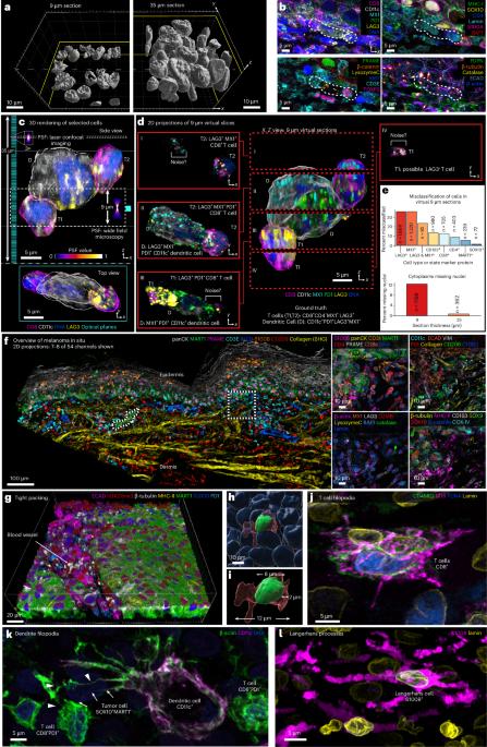
Diseases such as cancer involve alterations in cell proportions, states and interactions, as well as complex changes in tissue morphology and architecture. Histopathological diagnosis of disease and most multiplexed spatial profiling relies on inspecting thin (4–5 µm) specimens. Here we describe a high-plex cyclic immunofluorescence method for three-dimensional tissue imaging and use it to show that few, if any, cells are intact in conventional thin tissue sections, reducing the accuracy of cell phenotyping and interaction analysis. However, three-dimensional cyclic immunofluorescence of sections eightfold to tenfold thicker enables accurate morphological assessment of diverse protein markers in intact tumor, immune and stromal cells. Moreover, the high resolution of this confocal approach generates images of cells in a preserved tissue environment at a level of detail previously limited to cell culture. Precise imaging of cell membranes also makes it possible to detect and map cell–cell contacts and juxtracrine signaling complexes in immune cell niches. Confocal microscopy enables high-resolution, high-plex 3D cyclic immunofluorescence of 30- to 50-µm-thick tissue sections. The approach allows for rich phenotypic assessments of intact cells and intercellular interactions with subcellular resolution.
人类肿瘤细胞状态和免疫龛的高度多路3D分析。
癌症等疾病涉及细胞比例、状态和相互作用的改变,以及组织形态和结构的复杂变化。疾病的组织病理学诊断和大多数多重空间分析依赖于检查薄(4-5µm)的标本。在这里,我们描述了一种用于三维组织成像的高plex循环免疫荧光方法,并使用它来显示很少(如果有的话)细胞在常规薄组织切片中是完整的,从而降低了细胞表型和相互作用分析的准确性。然而,厚8至10倍切片的三维循环免疫荧光能够准确地评估完整肿瘤、免疫细胞和基质细胞中的各种蛋白质标记物。此外,这种共聚焦方法的高分辨率产生的细胞图像在一个保存的组织环境中的细节水平以前仅限于细胞培养。细胞膜的精确成像也使得检测和绘制免疫细胞壁龛中的细胞接触和近线信号复合物成为可能。
本文章由计算机程序翻译,如有差异,请以英文原文为准。
求助全文
约1分钟内获得全文
求助全文
来源期刊
Nature Methods
生物-生化研究方法
CiteScore
58.70
自引率
1.70%
发文量
326
审稿时长
1 months
期刊介绍:
Nature Methods is a monthly journal that focuses on publishing innovative methods and substantial enhancements to fundamental life sciences research techniques. Geared towards a diverse, interdisciplinary readership of researchers in academia and industry engaged in laboratory work, the journal offers new tools for research and emphasizes the immediate practical significance of the featured work. It publishes primary research papers and reviews recent technical and methodological advancements, with a particular interest in primary methods papers relevant to the biological and biomedical sciences. This includes methods rooted in chemistry with practical applications for studying biological problems.
 求助内容:
求助内容: 应助结果提醒方式:


