Uma Devaraj, Chitra Veluthat, Kavitha Venkatnarayan, Uma Maheswari Krishnaswamy, Priya Ramachandran
{"title":"Comparison of Lung Ultrasound Findings in Patients with Pulmonary Tuberculosis and Lobar Pneumonia: A Case-control Study.","authors":"Uma Devaraj, Chitra Veluthat, Kavitha Venkatnarayan, Uma Maheswari Krishnaswamy, Priya Ramachandran","doi":"10.4103/jmu.jmu_52_24","DOIUrl":null,"url":null,"abstract":"<p><strong>Background: </strong>The utility of lung ultrasound (LUS) in diagnosing respiratory disorders is being studied only in recent times. We aimed to describe the ultrasound (USG) features of pulmonary tuberculosis (TB) and compare them with those of lobar pneumonia. In addition, the LUS findings of both diseases were corroborated with chest X-ray findings.</p><p><strong>Methods: </strong>The study subjects consisted of adult subjects recently diagnosed with pulmonary TB and those diagnosed with lobar pneumonia. Both subsets of patients underwent LUS evaluation.</p><p><strong>Results: </strong>Ninety-six subjects with 64 microbiologically confirmed TB and 32 lobar pneumonia patients were included. The study subjects' mean age was 46.78 ± 15.75 years and the majority were males (<i>n</i> = 62; 64.6%). LUS showed focal interstitial pattern, cavity, and irregular pleura in TB patients which were significantly different (<i>P</i> ≤ 0.001) from the findings of air bronchogram and/or shred sign seen in patients with lobar pneumonia. The overall sensitivity of LUS compared to X-ray, to identify abnormalities in TB and lobar pneumonia patients, was 88.6%. The LUS and CXR findings were concordant in 93.75% of TB patients and 90.6%) of lobar pneumonia patients. Additional USG abnormalities other than that seen on CXR were demonstrated in 13 (20.3%) TB patients.</p><p><strong>Conclusion: </strong>LUS is a valuable tool to detect TB and lobar pneumonia and can discriminate between the two conditions. LUS performance was on par with CXR in the detection of abnormalities. The lack of radiation exposure and portability of LUS makes it an attractive tool for bedside use as well as in field conditions where radiography may not be readily available.</p>","PeriodicalId":45466,"journal":{"name":"Journal of Medical Ultrasound","volume":"33 3","pages":"216-221"},"PeriodicalIF":0.8000,"publicationDate":"2025-03-21","publicationTypes":"Journal Article","fieldsOfStudy":null,"isOpenAccess":false,"openAccessPdf":"https://www.ncbi.nlm.nih.gov/pmc/articles/PMC12463366/pdf/","citationCount":"0","resultStr":null,"platform":"Semanticscholar","paperid":null,"PeriodicalName":"Journal of Medical Ultrasound","FirstCategoryId":"1085","ListUrlMain":"https://doi.org/10.4103/jmu.jmu_52_24","RegionNum":0,"RegionCategory":null,"ArticlePicture":[],"TitleCN":null,"AbstractTextCN":null,"PMCID":null,"EPubDate":"2025/7/1 0:00:00","PubModel":"eCollection","JCR":"Q4","JCRName":"RADIOLOGY, NUCLEAR MEDICINE & MEDICAL IMAGING","Score":null,"Total":0}
引用次数: 0
Abstract
Background: The utility of lung ultrasound (LUS) in diagnosing respiratory disorders is being studied only in recent times. We aimed to describe the ultrasound (USG) features of pulmonary tuberculosis (TB) and compare them with those of lobar pneumonia. In addition, the LUS findings of both diseases were corroborated with chest X-ray findings.
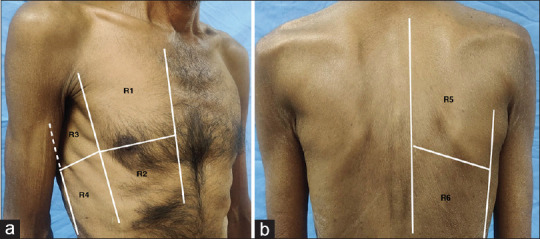
Methods: The study subjects consisted of adult subjects recently diagnosed with pulmonary TB and those diagnosed with lobar pneumonia. Both subsets of patients underwent LUS evaluation.
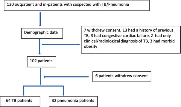
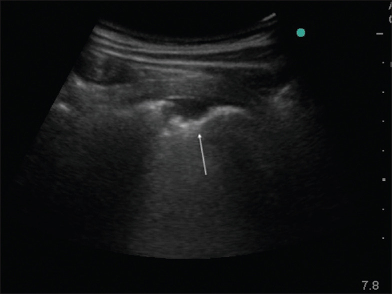
Results: Ninety-six subjects with 64 microbiologically confirmed TB and 32 lobar pneumonia patients were included. The study subjects' mean age was 46.78 ± 15.75 years and the majority were males (n = 62; 64.6%). LUS showed focal interstitial pattern, cavity, and irregular pleura in TB patients which were significantly different (P ≤ 0.001) from the findings of air bronchogram and/or shred sign seen in patients with lobar pneumonia. The overall sensitivity of LUS compared to X-ray, to identify abnormalities in TB and lobar pneumonia patients, was 88.6%. The LUS and CXR findings were concordant in 93.75% of TB patients and 90.6%) of lobar pneumonia patients. Additional USG abnormalities other than that seen on CXR were demonstrated in 13 (20.3%) TB patients.
Conclusion: LUS is a valuable tool to detect TB and lobar pneumonia and can discriminate between the two conditions. LUS performance was on par with CXR in the detection of abnormalities. The lack of radiation exposure and portability of LUS makes it an attractive tool for bedside use as well as in field conditions where radiography may not be readily available.
期刊介绍:
The Journal of Medical Ultrasound is the peer-reviewed publication of the Asian Federation of Societies for Ultrasound in Medicine and Biology, and the Chinese Taipei Society of Ultrasound in Medicine. Its aim is to promote clinical and scientific research in ultrasonography, and to serve as a channel of communication among sonologists, sonographers, and medical ultrasound physicians in the Asia-Pacific region and wider international community. The Journal invites original contributions relating to the clinical and laboratory investigations and applications of ultrasonography.

 求助内容:
求助内容: 应助结果提醒方式:


