Hatem Saadeldin Mohammed, Yasser A Elmotaleb Gazar, Saad Ghanem, Doaa Waseem Nada, Ahmed Maaty, Adel Ibrahim Azzam
{"title":"Ultrasonographic Evaluation of the Distal Medial Hamstring Tendons and their Association with Posteromedial Knee Pain.","authors":"Hatem Saadeldin Mohammed, Yasser A Elmotaleb Gazar, Saad Ghanem, Doaa Waseem Nada, Ahmed Maaty, Adel Ibrahim Azzam","doi":"10.4103/jmu.jmu_56_24","DOIUrl":null,"url":null,"abstract":"<p><strong>Background: </strong>Periarticular abnormalities are common ultrasonographic (U/S) findings in individuals with knee pain. Incidental U/S observations, including thickening of the distal hamstring tendons, require explanations for their clinical importance. In addition, it is unclear whether or not these tendon modifications are related to knee pain. The objective is to determine U/S findings of distal medial hamstring tendons in patients with posteromedial (PM) knee pain and assess the diagnostic significance of tendon thickness in predicting tendinopathy in those patients.</p><p><strong>Methods: </strong>We studied the distal medial hamstring tendons (semimembranosus [SM] and semitendinosus [ST]) of 104 patients (104 knees) with nontraumatic unilateral PM knee pain and 118 healthy controls (236 knees). U/S evaluations included tendon thickness, echogenicity, the presence of intrasubstance tears, calcifications, and vascularity.</p><p><strong>Results: </strong>The mean age of patients and controls was 51.7 ± 10.4 years and 49.8 ± 9.9 years, respectively. The mean Visual Analog Scale (VAS) for pain among patients was 5.1, with 58.6% of them reporting pain at the medial joint line. The study patients had significantly higher mean SM and ST tendon thicknesses than the controls (7.17 mm vs. 5.46 mm and 3.93 mm vs. 3.45 mm, respectively). U/S abnormalities among patients were hypoechogenicity (62.5%), intrasubstance tears (31.7%), loss of fibrillar pattern (23.1%), baker cyst (20.2%), calcification (18.3%), anserine bursitis (11.5%), and neovascularization (6.7%). We found significant correlations between tendon thickness and VAS (<i>r</i> = 0.752, <i>P</i> = 0.004) as well as pain location (<i>r</i> = 0.680, <i>P</i> = 0.008). SM tendon thickness measured by U/S was more accurate in predicting tendinopathy than ST (80.6% vs. 68.9%).</p><p><strong>Conclusion: </strong>U/S changes tend to occur frequently in individuals experiencing PM knee pain. Among the various abnormalities detectable by U/S, an increase in tendon thickness serves as a reliable indicator of tendinopathy and correlates strongly with the location and severity of knee pain. When dealing with PM knee pain, a comprehensive evaluation of the distal medial hamstring tendons through U/S examination can be instrumental in achieving a timely and accurate diagnosis as well as an effective treatment plan.</p>","PeriodicalId":45466,"journal":{"name":"Journal of Medical Ultrasound","volume":"33 3","pages":"241-247"},"PeriodicalIF":0.8000,"publicationDate":"2025-03-10","publicationTypes":"Journal Article","fieldsOfStudy":null,"isOpenAccess":false,"openAccessPdf":"https://www.ncbi.nlm.nih.gov/pmc/articles/PMC12463360/pdf/","citationCount":"0","resultStr":null,"platform":"Semanticscholar","paperid":null,"PeriodicalName":"Journal of Medical Ultrasound","FirstCategoryId":"1085","ListUrlMain":"https://doi.org/10.4103/jmu.jmu_56_24","RegionNum":0,"RegionCategory":null,"ArticlePicture":[],"TitleCN":null,"AbstractTextCN":null,"PMCID":null,"EPubDate":"2025/7/1 0:00:00","PubModel":"eCollection","JCR":"Q4","JCRName":"RADIOLOGY, NUCLEAR MEDICINE & MEDICAL IMAGING","Score":null,"Total":0}
引用次数: 0
Abstract
Background: Periarticular abnormalities are common ultrasonographic (U/S) findings in individuals with knee pain. Incidental U/S observations, including thickening of the distal hamstring tendons, require explanations for their clinical importance. In addition, it is unclear whether or not these tendon modifications are related to knee pain. The objective is to determine U/S findings of distal medial hamstring tendons in patients with posteromedial (PM) knee pain and assess the diagnostic significance of tendon thickness in predicting tendinopathy in those patients.
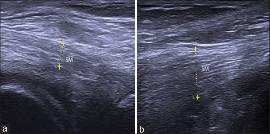
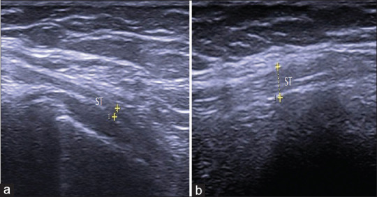
Methods: We studied the distal medial hamstring tendons (semimembranosus [SM] and semitendinosus [ST]) of 104 patients (104 knees) with nontraumatic unilateral PM knee pain and 118 healthy controls (236 knees). U/S evaluations included tendon thickness, echogenicity, the presence of intrasubstance tears, calcifications, and vascularity.
Results: The mean age of patients and controls was 51.7 ± 10.4 years and 49.8 ± 9.9 years, respectively. The mean Visual Analog Scale (VAS) for pain among patients was 5.1, with 58.6% of them reporting pain at the medial joint line. The study patients had significantly higher mean SM and ST tendon thicknesses than the controls (7.17 mm vs. 5.46 mm and 3.93 mm vs. 3.45 mm, respectively). U/S abnormalities among patients were hypoechogenicity (62.5%), intrasubstance tears (31.7%), loss of fibrillar pattern (23.1%), baker cyst (20.2%), calcification (18.3%), anserine bursitis (11.5%), and neovascularization (6.7%). We found significant correlations between tendon thickness and VAS (r = 0.752, P = 0.004) as well as pain location (r = 0.680, P = 0.008). SM tendon thickness measured by U/S was more accurate in predicting tendinopathy than ST (80.6% vs. 68.9%).
Conclusion: U/S changes tend to occur frequently in individuals experiencing PM knee pain. Among the various abnormalities detectable by U/S, an increase in tendon thickness serves as a reliable indicator of tendinopathy and correlates strongly with the location and severity of knee pain. When dealing with PM knee pain, a comprehensive evaluation of the distal medial hamstring tendons through U/S examination can be instrumental in achieving a timely and accurate diagnosis as well as an effective treatment plan.
期刊介绍:
The Journal of Medical Ultrasound is the peer-reviewed publication of the Asian Federation of Societies for Ultrasound in Medicine and Biology, and the Chinese Taipei Society of Ultrasound in Medicine. Its aim is to promote clinical and scientific research in ultrasonography, and to serve as a channel of communication among sonologists, sonographers, and medical ultrasound physicians in the Asia-Pacific region and wider international community. The Journal invites original contributions relating to the clinical and laboratory investigations and applications of ultrasonography.


 求助内容:
求助内容: 应助结果提醒方式:


