Mykhailo Nahorniak, Daniel Horák, Miroslav Šlouf, Miloš Steinhart, Oleksandr Shapoval, Hana Engstová and Petr Ježek
{"title":"Lanthanide-based UCNPs: toxicity evaluation and interaction of ultrasmall core vs. core–shell nanoparticles with cells","authors":"Mykhailo Nahorniak, Daniel Horák, Miroslav Šlouf, Miloš Steinhart, Oleksandr Shapoval, Hana Engstová and Petr Ježek","doi":"10.1039/D5MA00542F","DOIUrl":null,"url":null,"abstract":"<p >We describe a new concept for preparation of ultrasmall NaYF<small><sub>4</sub></small>:Yb,Er upconversion nanoparticles (UCNPs) with a diameter of 7 nm, depending on the amount of water added in the polymerization mixture, which affects the nucleation and growth of the particles. The morphology and structure of the nanoparticles were thoroughly characterized both in the dried state (TEM including elemental analysis and electron diffraction) and in solution (small and wide-angle X-ray scattering and dynamic light scattering). A thick NaYF<small><sub>4</sub></small> shell was subsequently introduced onto the particles, which significantly increased the luminescence by minimizing surface quenching effects and passivating the core from the surrounding environment. To make the particles dispersible in the aqueous environment natural for biological applications, they were coated with a ∼6 nm thick hydrophilic silica layer. This increased the size of core and core–shell UCNPs to 20 and ∼50 nm. All the developed particles exhibited non-cytotoxicity tested in insulinoma INS-1E cells. The upconversion luminescence of these nanoparticles incubated with INS-1E cells showed a similar pattern to that of the particles themselves. The small biocompatible UCNPs developed in this study are promising candidates for non-invasive and non-destructive applications in bioimaging. Thanks to their advantageous properties, <em>i.e.</em>, small size, adjustable optical properties and ability to interact with and easily penetrate cells, they are suitable for future use in platforms for targeted drug delivery and advanced diagnostic technologies.</p>","PeriodicalId":18242,"journal":{"name":"Materials Advances","volume":" 19","pages":" 6907-6918"},"PeriodicalIF":4.7000,"publicationDate":"2025-08-25","publicationTypes":"Journal Article","fieldsOfStudy":null,"isOpenAccess":false,"openAccessPdf":"https://pubs.rsc.org/en/content/articlepdf/2025/ma/d5ma00542f?page=search","citationCount":"0","resultStr":null,"platform":"Semanticscholar","paperid":null,"PeriodicalName":"Materials Advances","FirstCategoryId":"1085","ListUrlMain":"https://pubs.rsc.org/en/content/articlelanding/2025/ma/d5ma00542f","RegionNum":0,"RegionCategory":null,"ArticlePicture":[],"TitleCN":null,"AbstractTextCN":null,"PMCID":null,"EPubDate":"","PubModel":"","JCR":"Q2","JCRName":"MATERIALS SCIENCE, MULTIDISCIPLINARY","Score":null,"Total":0}
引用次数: 0
Abstract
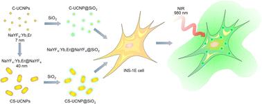
We describe a new concept for preparation of ultrasmall NaYF4:Yb,Er upconversion nanoparticles (UCNPs) with a diameter of 7 nm, depending on the amount of water added in the polymerization mixture, which affects the nucleation and growth of the particles. The morphology and structure of the nanoparticles were thoroughly characterized both in the dried state (TEM including elemental analysis and electron diffraction) and in solution (small and wide-angle X-ray scattering and dynamic light scattering). A thick NaYF4 shell was subsequently introduced onto the particles, which significantly increased the luminescence by minimizing surface quenching effects and passivating the core from the surrounding environment. To make the particles dispersible in the aqueous environment natural for biological applications, they were coated with a ∼6 nm thick hydrophilic silica layer. This increased the size of core and core–shell UCNPs to 20 and ∼50 nm. All the developed particles exhibited non-cytotoxicity tested in insulinoma INS-1E cells. The upconversion luminescence of these nanoparticles incubated with INS-1E cells showed a similar pattern to that of the particles themselves. The small biocompatible UCNPs developed in this study are promising candidates for non-invasive and non-destructive applications in bioimaging. Thanks to their advantageous properties, i.e., small size, adjustable optical properties and ability to interact with and easily penetrate cells, they are suitable for future use in platforms for targeted drug delivery and advanced diagnostic technologies.

 求助内容:
求助内容: 应助结果提醒方式:


