A P Pershina-Miliutina, A K Eremkina, I D Ozhimalov, А V Khairieva, A M Gorbacheva, S V Ronzhina, N G Mokrysheva
{"title":"[Effect of preoperative bisphosphonate therapy on bone mineral density in patients with primary hyperparathyroidism one year after parathyroidectomy].","authors":"A P Pershina-Miliutina, A K Eremkina, I D Ozhimalov, А V Khairieva, A M Gorbacheva, S V Ronzhina, N G Mokrysheva","doi":"10.14341/probl13574","DOIUrl":null,"url":null,"abstract":"<p><strong>Background: </strong>The main treatment for primary hyperparathyroidism (PHPT) is parathyroidectomy (PTE), conservative therapy, including bisphosphonates, can be used for preoperative correction of hypercalcemia, as well as to improve bone tissue condition among individuals for whom surgery should be postponed or cannot be performed due to high perioperative risks. The question of the effect of bisphosphonates on bone tissue after surgery remains open.</p><p><strong>Aim: </strong>To study the effect of preoperative bisphosphonate therapy on BMD parameters assessed in DXA and 3D-DXA in patients with PHPT one year after radical PTE.</p><p><strong>Materials and methods: </strong>The study was conducted on the basis of the Department of pathology of the parathyroid glands and disorders of mineral metabolism of \"Endocrinology Research Center\" state-funded research facility of the Ministry of Health of the Russian Federation. The study included 50 patients (2 men, 48 women), divided into two groups depending on the presence or absence of preoperative bisphosphonate (BF) therapy. The methods of DXA and 3D-DXA using 3D-Shaper Medical software were used to evaluate BMD and bone microarchitectonics. The statistical analysis was performed using the R language and the Statistica v.13 package.</p><p><strong>Results: </strong>At the time of the disease's manifestation, both groups were comparable in terms of the main indicators of calcium phosphorus metabolism, with the exception of the level of beta-crosslapse, which was higher in the group without preoperative BPh therapy (p<0,001). There were also no differences in the parameters of DXA and 3D-DXA. After surgery, both groups showed a comparable increase in BMD based on the results of DXA in the main parts of the skeleton and 3D-DXA in the femur. Changes at the level of the statistical trend were obtained for the 3D-DXA parameters, the final absolute values of which were slightly higher in the second group, including the thickness of the cortical layer in the femur as a whole and in the neck. When comparing the results of DXA before and after PTE in patients receiving BPh, statistically significant differences in absolute BMD values were obtained only in the lumbar spine (p<0,001).According to 3D-DXA data, statistically significant differences were found only in the volume of mineral density of the trabecular bone of the femur as a whole (p=0,001).When analyzing up to - in the second group, statistically significant differences in absolute BMD values were observed in the lumbar region (p<0,001), in the hip as a whole (p<0,001) and in its neck (p=0,001).According to 3D-DXA data, statistically significant differences were found in three of the eight analyzed indicators, the volume of mineral density of the trabecular bone of the femur as a whole and in the neck (p<0,001 for both), as well as the volume of mineral density of the cortical bone in the neck, (p=0,001).</p><p><strong>Conclusion: </strong>The 3D-DXA method allows us to evaluate not only BMD, but also its microarchitectonics, which is important for predicting the risk of fractures in patients with PHPT. Studies have shown that preoperative BPh therapy can negatively affect the recovery of BMD after PTE, especially in cortical bone tissue. Further studies are needed to confirm these data and clarify the effect of CF on the postoperative course of PHPT.</p>","PeriodicalId":101419,"journal":{"name":"Problemy endokrinologii","volume":"71 4","pages":"57-70"},"PeriodicalIF":0.0000,"publicationDate":"2025-09-14","publicationTypes":"Journal Article","fieldsOfStudy":null,"isOpenAccess":false,"openAccessPdf":"https://www.ncbi.nlm.nih.gov/pmc/articles/PMC12489962/pdf/","citationCount":"0","resultStr":null,"platform":"Semanticscholar","paperid":null,"PeriodicalName":"Problemy endokrinologii","FirstCategoryId":"1085","ListUrlMain":"https://doi.org/10.14341/probl13574","RegionNum":0,"RegionCategory":null,"ArticlePicture":[],"TitleCN":null,"AbstractTextCN":null,"PMCID":null,"EPubDate":"","PubModel":"","JCR":"","JCRName":"","Score":null,"Total":0}
引用次数: 0
Abstract
Background: The main treatment for primary hyperparathyroidism (PHPT) is parathyroidectomy (PTE), conservative therapy, including bisphosphonates, can be used for preoperative correction of hypercalcemia, as well as to improve bone tissue condition among individuals for whom surgery should be postponed or cannot be performed due to high perioperative risks. The question of the effect of bisphosphonates on bone tissue after surgery remains open.
Aim: To study the effect of preoperative bisphosphonate therapy on BMD parameters assessed in DXA and 3D-DXA in patients with PHPT one year after radical PTE.
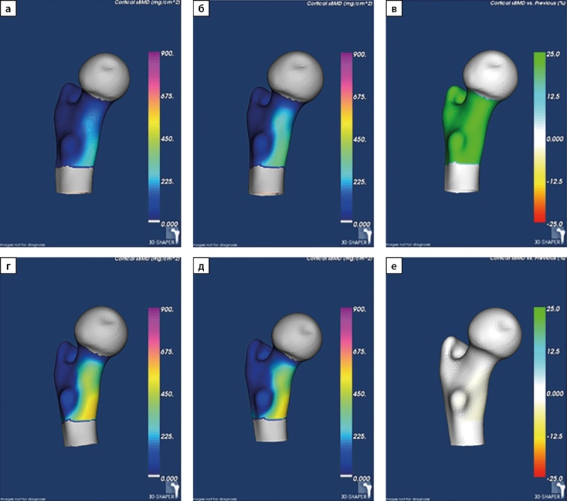
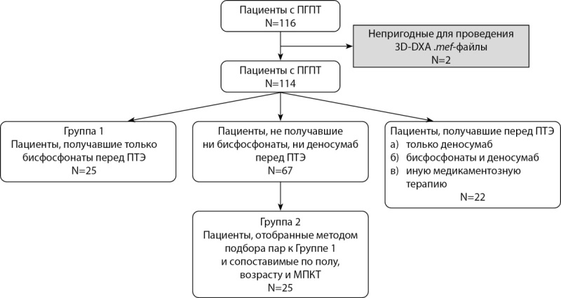
Materials and methods: The study was conducted on the basis of the Department of pathology of the parathyroid glands and disorders of mineral metabolism of "Endocrinology Research Center" state-funded research facility of the Ministry of Health of the Russian Federation. The study included 50 patients (2 men, 48 women), divided into two groups depending on the presence or absence of preoperative bisphosphonate (BF) therapy. The methods of DXA and 3D-DXA using 3D-Shaper Medical software were used to evaluate BMD and bone microarchitectonics. The statistical analysis was performed using the R language and the Statistica v.13 package.
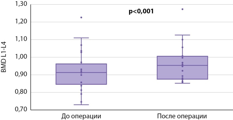
Results: At the time of the disease's manifestation, both groups were comparable in terms of the main indicators of calcium phosphorus metabolism, with the exception of the level of beta-crosslapse, which was higher in the group without preoperative BPh therapy (p<0,001). There were also no differences in the parameters of DXA and 3D-DXA. After surgery, both groups showed a comparable increase in BMD based on the results of DXA in the main parts of the skeleton and 3D-DXA in the femur. Changes at the level of the statistical trend were obtained for the 3D-DXA parameters, the final absolute values of which were slightly higher in the second group, including the thickness of the cortical layer in the femur as a whole and in the neck. When comparing the results of DXA before and after PTE in patients receiving BPh, statistically significant differences in absolute BMD values were obtained only in the lumbar spine (p<0,001).According to 3D-DXA data, statistically significant differences were found only in the volume of mineral density of the trabecular bone of the femur as a whole (p=0,001).When analyzing up to - in the second group, statistically significant differences in absolute BMD values were observed in the lumbar region (p<0,001), in the hip as a whole (p<0,001) and in its neck (p=0,001).According to 3D-DXA data, statistically significant differences were found in three of the eight analyzed indicators, the volume of mineral density of the trabecular bone of the femur as a whole and in the neck (p<0,001 for both), as well as the volume of mineral density of the cortical bone in the neck, (p=0,001).
Conclusion: The 3D-DXA method allows us to evaluate not only BMD, but also its microarchitectonics, which is important for predicting the risk of fractures in patients with PHPT. Studies have shown that preoperative BPh therapy can negatively affect the recovery of BMD after PTE, especially in cortical bone tissue. Further studies are needed to confirm these data and clarify the effect of CF on the postoperative course of PHPT.

 求助内容:
求助内容: 应助结果提醒方式:


