A rare case report of esophageal neuroendocrine carcinoma mimicking a submucosal tumor: Diagnosis confirmed by endoscopic ultrasound-guided fine-needle aspiration.
Chunxiao Hu, Di Zhu, Ping Li, Xiaohua Ye, Zhiyi Chen
{"title":"A rare case report of esophageal neuroendocrine carcinoma mimicking a submucosal tumor: Diagnosis confirmed by endoscopic ultrasound-guided fine-needle aspiration.","authors":"Chunxiao Hu, Di Zhu, Ping Li, Xiaohua Ye, Zhiyi Chen","doi":"10.1177/03000605251381453","DOIUrl":null,"url":null,"abstract":"<p><p>Esophageal neuroendocrine carcinoma is an exceedingly rare and aggressive malignancy, accounting for less than 1% of all esophageal cancers. This case report presents a unique instance of esophageal neuroendocrine carcinoma that initially manifests as a submucosal tumor-like lesion, an atypical and under-recognized presentation that can mimic benign conditions such as leiomyomas or gastrointestinal stromal tumors. Conventional endoscopic biopsy, often the first-line diagnostic tool, failed to provide a definitive diagnosis due to the tumor's deep submucosal location and intact overlying mucosa. However, the utilization of endoscopic ultrasound-guided fine-needle aspiration proved instrumental in obtaining sufficient tissue for histopathological and immunohistochemical analyses, confirming the diagnosis of esophageal neuroendocrine carcinoma. This case underscores the critical role of endoscopic ultrasound-guided fine-needle aspiration in diagnosing rare esophageal malignancies that evade standard diagnostic approaches. Furthermore, it emphasizes the importance of considering esophageal neuroendocrine carcinoma in the differential diagnosis of unusual esophageal lesions, particularly when they exhibit unusual features or fail to respond to conventional management. Early and accurate diagnosis is essential for guiding appropriate therapeutic strategies and improving patient outcomes.</p>","PeriodicalId":16129,"journal":{"name":"Journal of International Medical Research","volume":"53 9","pages":"3000605251381453"},"PeriodicalIF":1.5000,"publicationDate":"2025-09-01","publicationTypes":"Journal Article","fieldsOfStudy":null,"isOpenAccess":false,"openAccessPdf":"https://www.ncbi.nlm.nih.gov/pmc/articles/PMC12475813/pdf/","citationCount":"0","resultStr":null,"platform":"Semanticscholar","paperid":null,"PeriodicalName":"Journal of International Medical Research","FirstCategoryId":"3","ListUrlMain":"https://doi.org/10.1177/03000605251381453","RegionNum":4,"RegionCategory":"医学","ArticlePicture":[],"TitleCN":null,"AbstractTextCN":null,"PMCID":null,"EPubDate":"2025/9/26 0:00:00","PubModel":"Epub","JCR":"Q4","JCRName":"MEDICINE, RESEARCH & EXPERIMENTAL","Score":null,"Total":0}
引用次数: 0
Abstract
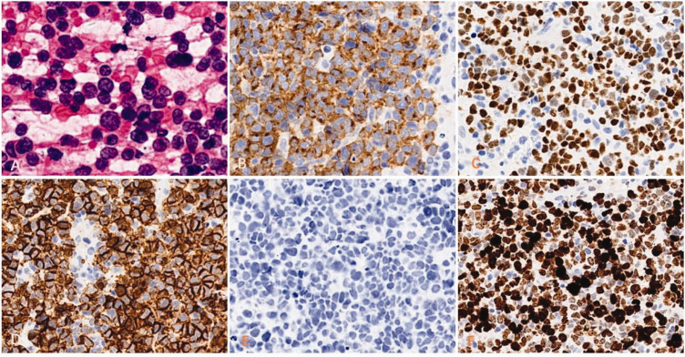
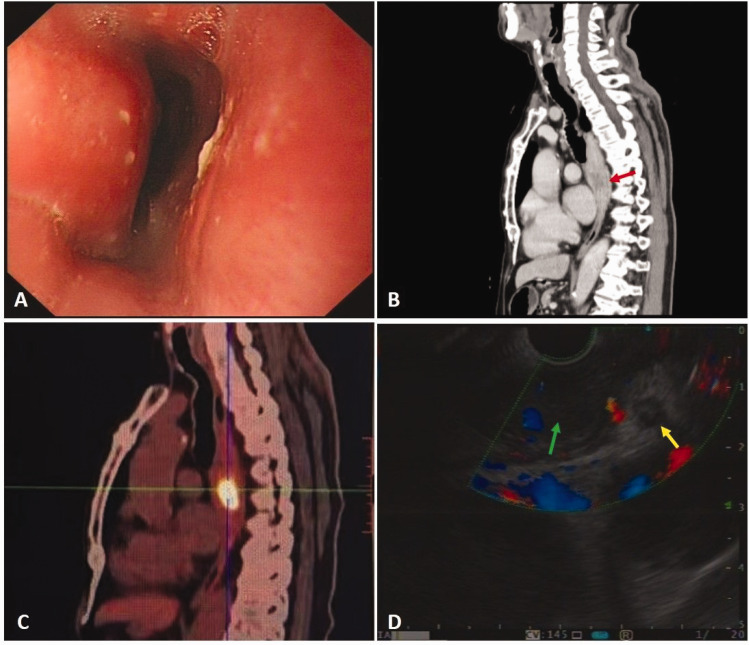
Esophageal neuroendocrine carcinoma is an exceedingly rare and aggressive malignancy, accounting for less than 1% of all esophageal cancers. This case report presents a unique instance of esophageal neuroendocrine carcinoma that initially manifests as a submucosal tumor-like lesion, an atypical and under-recognized presentation that can mimic benign conditions such as leiomyomas or gastrointestinal stromal tumors. Conventional endoscopic biopsy, often the first-line diagnostic tool, failed to provide a definitive diagnosis due to the tumor's deep submucosal location and intact overlying mucosa. However, the utilization of endoscopic ultrasound-guided fine-needle aspiration proved instrumental in obtaining sufficient tissue for histopathological and immunohistochemical analyses, confirming the diagnosis of esophageal neuroendocrine carcinoma. This case underscores the critical role of endoscopic ultrasound-guided fine-needle aspiration in diagnosing rare esophageal malignancies that evade standard diagnostic approaches. Furthermore, it emphasizes the importance of considering esophageal neuroendocrine carcinoma in the differential diagnosis of unusual esophageal lesions, particularly when they exhibit unusual features or fail to respond to conventional management. Early and accurate diagnosis is essential for guiding appropriate therapeutic strategies and improving patient outcomes.
期刊介绍:
_Journal of International Medical Research_ is a leading international journal for rapid publication of original medical, pre-clinical and clinical research, reviews, preliminary and pilot studies on a page charge basis.
As a service to authors, every article accepted by peer review will be given a full technical edit to make papers as accessible and readable to the international medical community as rapidly as possible.
Once the technical edit queries have been answered to the satisfaction of the journal, the paper will be published and made available freely to everyone under a creative commons licence.
Symposium proceedings, summaries of presentations or collections of medical, pre-clinical or clinical data on a specific topic are welcome for publication as supplements.
Print ISSN: 0300-0605
 求助内容:
求助内容: 应助结果提醒方式:


