Chong Ge, Yi Shi, Wei Wang, Anli Zhang, Mengqi Huang, Fang Zhao, Ao Li, Zhenzhong Feng, Minghui Wang, Haibo Wu
{"title":"Artificial Intelligence-driven image analysis for standardised programmed death-ligand 1 expression evaluation in non-small cell lung cancer.","authors":"Chong Ge, Yi Shi, Wei Wang, Anli Zhang, Mengqi Huang, Fang Zhao, Ao Li, Zhenzhong Feng, Minghui Wang, Haibo Wu","doi":"10.1186/s13000-025-01707-1","DOIUrl":null,"url":null,"abstract":"<p><strong>Background: </strong>Accurate assessment of programmed death-ligand 1 (PD-L1) immunohistochemical (IHC) expression is critical for immunotherapy in patients with non-small cell lung cancer (NSCLC). Yet, interpreting its staining is challenging, time-consuming, and causes inter-observer variability, potentially mis-stratifying patients. This necessitates the development of an artificial intelligence (AI) model to effectively quantify PD-L1 expression. Hence, we developed an AI-based deep-learning approach to automatically assess PD-L1 expression in NSCLC using IHC 22C3 assay-stained whole slide images (WSIs).</p><p><strong>Methods: </strong>A total of 706 patients with NSCLC were included in this study and 1212 WSIs were collected from three distinct study cohorts. We accurately matched the hematoxylin and eosin-stained images of the internal dataset with the IHC WSIs. Foreground regions containing tumor tissue were extracted from WSIs, and a multi-granular multiple-instance learning approach employing instance embeddings with coarse and fine granularities was implemented to extract patch-level morphological features. A multi-grained expression interpreter-based model aggregated these features to stratify PD-L1 expression status.</p><p><strong>Results: </strong>The model showed strong interpretive ability in all three cohorts and wide applicability to different specimen types. The macro-average area under the receiver operating characteristic curve (AUC) were 0.940/0.915/0.944 for surgical specimens, 0.955/0.844/0.865 for biopsy specimens, and 0.901/0.958/0.883 for metastases.</p><p><strong>Conclusion: </strong>This study emphasizes the potential benefits of deep learning in automatically, rapidly, and accurately inferring PD-L1 expression from complex IHC images. It also showcases how AI frameworks can improve routine digital pathology workflows in current PD-L1 detection methods.</p>","PeriodicalId":11237,"journal":{"name":"Diagnostic Pathology","volume":"20 1","pages":"106"},"PeriodicalIF":2.3000,"publicationDate":"2025-09-26","publicationTypes":"Journal Article","fieldsOfStudy":null,"isOpenAccess":false,"openAccessPdf":"https://www.ncbi.nlm.nih.gov/pmc/articles/PMC12465877/pdf/","citationCount":"0","resultStr":null,"platform":"Semanticscholar","paperid":null,"PeriodicalName":"Diagnostic Pathology","FirstCategoryId":"3","ListUrlMain":"https://doi.org/10.1186/s13000-025-01707-1","RegionNum":3,"RegionCategory":"医学","ArticlePicture":[],"TitleCN":null,"AbstractTextCN":null,"PMCID":null,"EPubDate":"","PubModel":"","JCR":"Q2","JCRName":"PATHOLOGY","Score":null,"Total":0}
引用次数: 0
Abstract
Background: Accurate assessment of programmed death-ligand 1 (PD-L1) immunohistochemical (IHC) expression is critical for immunotherapy in patients with non-small cell lung cancer (NSCLC). Yet, interpreting its staining is challenging, time-consuming, and causes inter-observer variability, potentially mis-stratifying patients. This necessitates the development of an artificial intelligence (AI) model to effectively quantify PD-L1 expression. Hence, we developed an AI-based deep-learning approach to automatically assess PD-L1 expression in NSCLC using IHC 22C3 assay-stained whole slide images (WSIs).
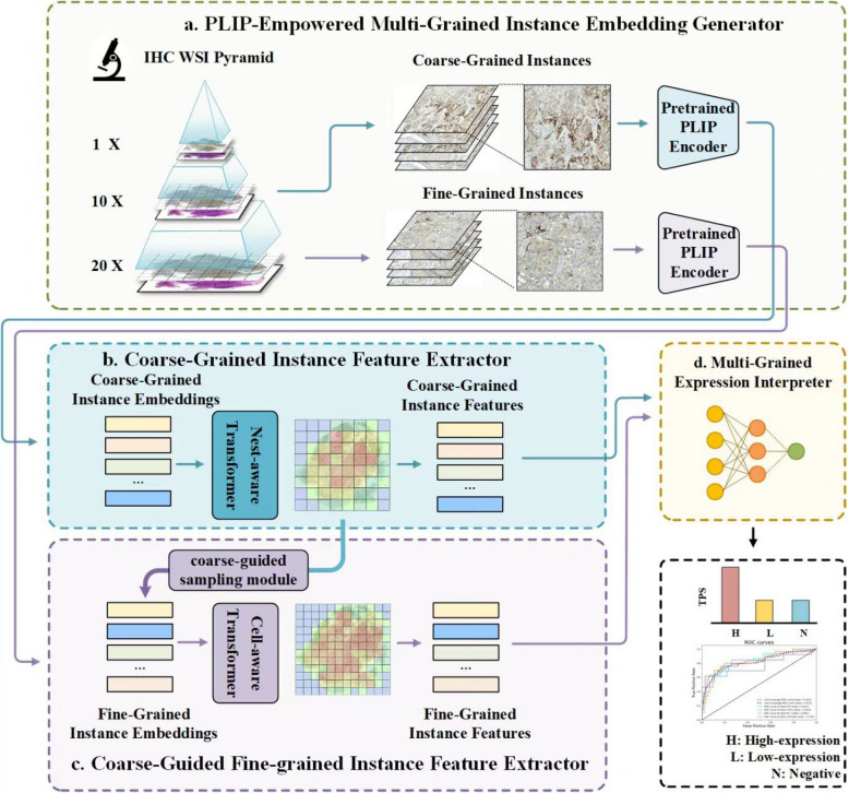
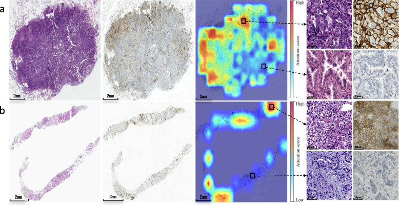
Methods: A total of 706 patients with NSCLC were included in this study and 1212 WSIs were collected from three distinct study cohorts. We accurately matched the hematoxylin and eosin-stained images of the internal dataset with the IHC WSIs. Foreground regions containing tumor tissue were extracted from WSIs, and a multi-granular multiple-instance learning approach employing instance embeddings with coarse and fine granularities was implemented to extract patch-level morphological features. A multi-grained expression interpreter-based model aggregated these features to stratify PD-L1 expression status.
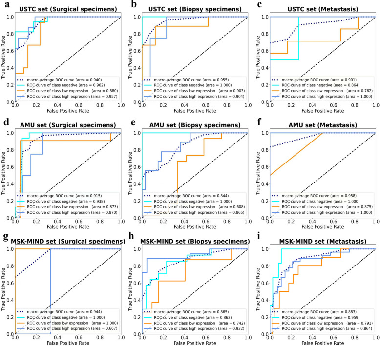
Results: The model showed strong interpretive ability in all three cohorts and wide applicability to different specimen types. The macro-average area under the receiver operating characteristic curve (AUC) were 0.940/0.915/0.944 for surgical specimens, 0.955/0.844/0.865 for biopsy specimens, and 0.901/0.958/0.883 for metastases.
Conclusion: This study emphasizes the potential benefits of deep learning in automatically, rapidly, and accurately inferring PD-L1 expression from complex IHC images. It also showcases how AI frameworks can improve routine digital pathology workflows in current PD-L1 detection methods.
期刊介绍:
Diagnostic Pathology is an open access, peer-reviewed, online journal that considers research in surgical and clinical pathology, immunology, and biology, with a special focus on cutting-edge approaches in diagnostic pathology and tissue-based therapy. The journal covers all aspects of surgical pathology, including classic diagnostic pathology, prognosis-related diagnosis (tumor stages, prognosis markers, such as MIB-percentage, hormone receptors, etc.), and therapy-related findings. The journal also focuses on the technological aspects of pathology, including molecular biology techniques, morphometry aspects (stereology, DNA analysis, syntactic structure analysis), communication aspects (telecommunication, virtual microscopy, virtual pathology institutions, etc.), and electronic education and quality assurance (for example interactive publication, on-line references with automated updating, etc.).

 求助内容:
求助内容: 应助结果提醒方式:


