Comparison of swept source - Optical coherence tomography angiography with fundus fluorescein angiography for detection of lesions in diabetic retinopathy.
Santosh Kumar Mahapatra, Anuja Mohanty, Amit Bidasaria, Anjalika Parhi
{"title":"Comparison of swept source - Optical coherence tomography angiography with fundus fluorescein angiography for detection of lesions in diabetic retinopathy.","authors":"Santosh Kumar Mahapatra, Anuja Mohanty, Amit Bidasaria, Anjalika Parhi","doi":"10.4103/tjo.TJO-D-24-00117","DOIUrl":null,"url":null,"abstract":"<p><strong>Purpose: </strong>The purpose of this study was to compare the rate of detection of diabetic retinopathy (DR) lesions and the agreement for grading DR severity between swept-source optical coherence tomography angiography (OCTA) and fundus fluorescein angiography (FFA) and establish the utility of OCTA as a noninvasive alternative to FFA.</p><p><strong>Materials and methods: </strong>116 eyes of 60 DR patients underwent OCTA with a 12 m × 12 mm acquisition protocol centered at the fovea followed by FFA. For each imaging technique, the presence or absence of DR lesions including microaneurysms, intraretinal microvascular abnormalities (IRMAs), new vessels on the disc (NVD), new vessels elsewhere (NVE), and nonperfusion areas (NPAs) was recorded. Statistical analysis was performed using IBM SPSS.22 using the McNemar test.</p><p><strong>Results: </strong>The detection rates were comparable in OCTA versus FFA for most DR lesions (<i>P</i> > 0.05) except microaneurysms (90 eyes, 77.6% in OCTA vs. 115 eyes, 99.1% in FFA). OCTA detected NPAs better than FFA (91 eyes, 78.5% vs. 78 eyes, 67.2%). There was an excellent agreement for the identification of IRMA (<i>κ</i> =0.791), NVD (<i>κ</i> =0.938), and NVE (<i>κ</i> =0.942); good agreement for the identification of NPA (<i>κ</i> =0.635) and poor agreement for microaneurysms (<i>κ</i> =0.058) identification. Overall, agreement in grading of DR severity between OCTA and FFA was good (<i>κ</i> =0.687).</p><p><strong>Conclusion: </strong>OCTA serves as a noninvasive, rapid imaging modality for evaluating retinal vascular changes in DR and can be the sole imaging modality in specific situations such as pregnancy, nephropathy, and in patients with uncontrolled diabetes and hypertension. OCTA is noninferior to FFA, and both modalities should be utilized as complementary imaging modalities to maximize their respective advantages and improve treatment outcomes.</p>","PeriodicalId":44978,"journal":{"name":"Taiwan Journal of Ophthalmology","volume":"15 3","pages":"443-449"},"PeriodicalIF":1.2000,"publicationDate":"2025-05-02","publicationTypes":"Journal Article","fieldsOfStudy":null,"isOpenAccess":false,"openAccessPdf":"https://www.ncbi.nlm.nih.gov/pmc/articles/PMC12456908/pdf/","citationCount":"0","resultStr":null,"platform":"Semanticscholar","paperid":null,"PeriodicalName":"Taiwan Journal of Ophthalmology","FirstCategoryId":"1085","ListUrlMain":"https://doi.org/10.4103/tjo.TJO-D-24-00117","RegionNum":0,"RegionCategory":null,"ArticlePicture":[],"TitleCN":null,"AbstractTextCN":null,"PMCID":null,"EPubDate":"2025/7/1 0:00:00","PubModel":"eCollection","JCR":"Q4","JCRName":"OPHTHALMOLOGY","Score":null,"Total":0}
引用次数: 0
Abstract
Purpose: The purpose of this study was to compare the rate of detection of diabetic retinopathy (DR) lesions and the agreement for grading DR severity between swept-source optical coherence tomography angiography (OCTA) and fundus fluorescein angiography (FFA) and establish the utility of OCTA as a noninvasive alternative to FFA.
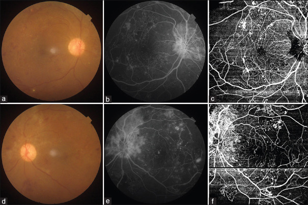
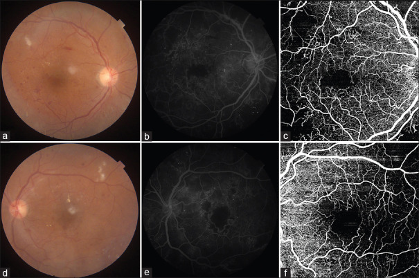
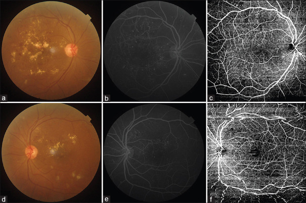
Materials and methods: 116 eyes of 60 DR patients underwent OCTA with a 12 m × 12 mm acquisition protocol centered at the fovea followed by FFA. For each imaging technique, the presence or absence of DR lesions including microaneurysms, intraretinal microvascular abnormalities (IRMAs), new vessels on the disc (NVD), new vessels elsewhere (NVE), and nonperfusion areas (NPAs) was recorded. Statistical analysis was performed using IBM SPSS.22 using the McNemar test.
Results: The detection rates were comparable in OCTA versus FFA for most DR lesions (P > 0.05) except microaneurysms (90 eyes, 77.6% in OCTA vs. 115 eyes, 99.1% in FFA). OCTA detected NPAs better than FFA (91 eyes, 78.5% vs. 78 eyes, 67.2%). There was an excellent agreement for the identification of IRMA (κ =0.791), NVD (κ =0.938), and NVE (κ =0.942); good agreement for the identification of NPA (κ =0.635) and poor agreement for microaneurysms (κ =0.058) identification. Overall, agreement in grading of DR severity between OCTA and FFA was good (κ =0.687).
Conclusion: OCTA serves as a noninvasive, rapid imaging modality for evaluating retinal vascular changes in DR and can be the sole imaging modality in specific situations such as pregnancy, nephropathy, and in patients with uncontrolled diabetes and hypertension. OCTA is noninferior to FFA, and both modalities should be utilized as complementary imaging modalities to maximize their respective advantages and improve treatment outcomes.

 求助内容:
求助内容: 应助结果提醒方式:


