{"title":"Applications of deep learning in the analysis of optical coherence tomography images for glaucoma-related diagnostics.","authors":"Kyle Bolo, Benjamin Y Xu","doi":"10.4103/tjo.TJO-D-24-00162","DOIUrl":null,"url":null,"abstract":"<p><p>Glaucoma is an optic neuropathy and the leading cause of irreversible blindness worldwide. Imaging of the ganglion cell complex and retinal nerve fiber layer with optical coherence tomography (OCT) is a noninvasive, high-resolution means of diagnosing and quantitatively monitoring glaucoma. In the anterior segment, OCT can also be used to assess the anterior chamber angle and identify angle closure, a risk factor for glaucoma. The interpretation of OCT images for accurate diagnosis requires expert-level knowledge of both the technology and glaucoma. Deep learning (DL) is a subfield of artificial intelligence (AI), which is gaining prominence in health care for its ability to interpret images and approximate clinician judgment. This review summarizes recent research that demonstrates how DL can contribute to the analysis of OCT images in glaucoma. Deep neural networks can assist clinicians in checking the quality of OCT scans, quantifying the thickness of optic nerve tissues, evaluating the anterior chamber angle, diagnosing glaucoma, and detecting the progression of existing glaucoma. As further work expands on the generalizability, equity, and explainability of these DL techniques, AI-driven clinical support tools may become available for glaucoma diagnostics.</p>","PeriodicalId":44978,"journal":{"name":"Taiwan Journal of Ophthalmology","volume":"15 3","pages":"354-363"},"PeriodicalIF":1.2000,"publicationDate":"2025-07-18","publicationTypes":"Journal Article","fieldsOfStudy":null,"isOpenAccess":false,"openAccessPdf":"https://www.ncbi.nlm.nih.gov/pmc/articles/PMC12456913/pdf/","citationCount":"0","resultStr":null,"platform":"Semanticscholar","paperid":null,"PeriodicalName":"Taiwan Journal of Ophthalmology","FirstCategoryId":"1085","ListUrlMain":"https://doi.org/10.4103/tjo.TJO-D-24-00162","RegionNum":0,"RegionCategory":null,"ArticlePicture":[],"TitleCN":null,"AbstractTextCN":null,"PMCID":null,"EPubDate":"2025/7/1 0:00:00","PubModel":"eCollection","JCR":"Q4","JCRName":"OPHTHALMOLOGY","Score":null,"Total":0}
引用次数: 0
Abstract
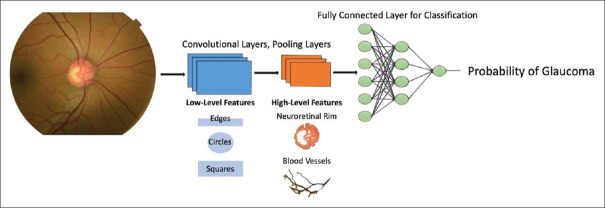
Glaucoma is an optic neuropathy and the leading cause of irreversible blindness worldwide. Imaging of the ganglion cell complex and retinal nerve fiber layer with optical coherence tomography (OCT) is a noninvasive, high-resolution means of diagnosing and quantitatively monitoring glaucoma. In the anterior segment, OCT can also be used to assess the anterior chamber angle and identify angle closure, a risk factor for glaucoma. The interpretation of OCT images for accurate diagnosis requires expert-level knowledge of both the technology and glaucoma. Deep learning (DL) is a subfield of artificial intelligence (AI), which is gaining prominence in health care for its ability to interpret images and approximate clinician judgment. This review summarizes recent research that demonstrates how DL can contribute to the analysis of OCT images in glaucoma. Deep neural networks can assist clinicians in checking the quality of OCT scans, quantifying the thickness of optic nerve tissues, evaluating the anterior chamber angle, diagnosing glaucoma, and detecting the progression of existing glaucoma. As further work expands on the generalizability, equity, and explainability of these DL techniques, AI-driven clinical support tools may become available for glaucoma diagnostics.

 求助内容:
求助内容: 应助结果提醒方式:


