Qingyu Chen, Stephanie Lauren Nolen, Sydni Adriana Spencer, Ji Yi
{"title":"The path to clinical translation for visible light optical coherence tomography in retinal imaging.","authors":"Qingyu Chen, Stephanie Lauren Nolen, Sydni Adriana Spencer, Ji Yi","doi":"10.4103/tjo.TJO-D-25-00078","DOIUrl":null,"url":null,"abstract":"<p><p>Visible light optical coherence tomography (VIS-OCT) has made significant progress in the past decade from <i>in vivo</i> proof-of-concept retinal imaging in preclinical models to human clinical translation. The technical advances of VIS-OCT imaging devices include new light sources, optical fiber components, balanced detection methods, and an array of data processing methods. We summarize the unique features of using VIS-OCT in comparison with near-infrared OCT (NIR-OCT), including ultra-high resolution, retinal microvascular oximetry, and reflectance spectroscopy. The ultra-high resolution is granted by the shorter wavelengths in the visible light range ~500-650 nm, as compared with the conventional OCT wavelengths >800 nm. Detailed sub-bandings in the inner plexiform layer and outer segment of photoreceptors, as well as in the retinal pigment epithelium and Bruch's membrane, are consistently resolved in VIS-OCT. The three-dimensional resolving capacity of VIS-OCT allows better isolation of hemoglobin absorption features, allowing blood oxygen saturation (SO<sub>2</sub>) calculation in retinal microvasculature. Oximetry calculations were performed down to the capillary level in humans, albeit through massive averaging, which was unattainable by previous methods. Advancing VIS-OCT technology has a high potential to produce significant clinical impact in ophthalmology in the near future.</p>","PeriodicalId":44978,"journal":{"name":"Taiwan Journal of Ophthalmology","volume":"15 3","pages":"389-398"},"PeriodicalIF":1.2000,"publicationDate":"2025-08-29","publicationTypes":"Journal Article","fieldsOfStudy":null,"isOpenAccess":false,"openAccessPdf":"https://www.ncbi.nlm.nih.gov/pmc/articles/PMC12456912/pdf/","citationCount":"0","resultStr":null,"platform":"Semanticscholar","paperid":null,"PeriodicalName":"Taiwan Journal of Ophthalmology","FirstCategoryId":"1085","ListUrlMain":"https://doi.org/10.4103/tjo.TJO-D-25-00078","RegionNum":0,"RegionCategory":null,"ArticlePicture":[],"TitleCN":null,"AbstractTextCN":null,"PMCID":null,"EPubDate":"2025/7/1 0:00:00","PubModel":"eCollection","JCR":"Q4","JCRName":"OPHTHALMOLOGY","Score":null,"Total":0}
引用次数: 0
Abstract
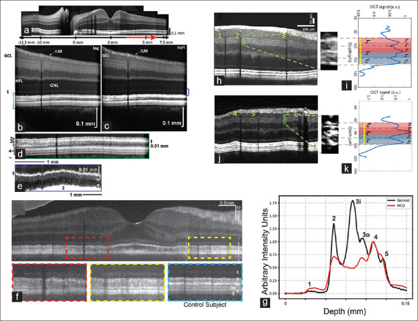
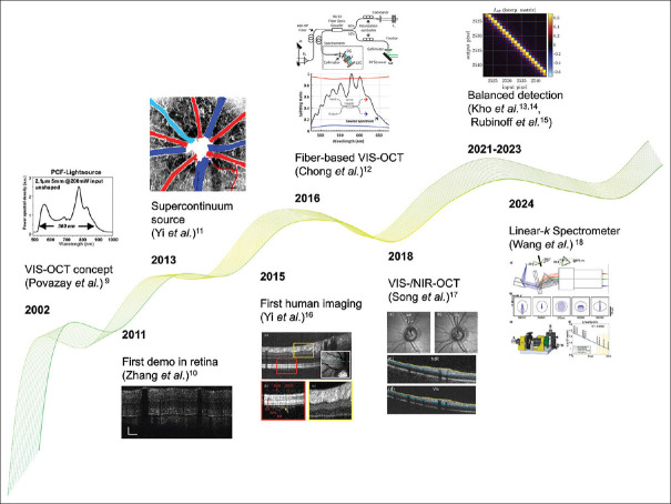
Visible light optical coherence tomography (VIS-OCT) has made significant progress in the past decade from in vivo proof-of-concept retinal imaging in preclinical models to human clinical translation. The technical advances of VIS-OCT imaging devices include new light sources, optical fiber components, balanced detection methods, and an array of data processing methods. We summarize the unique features of using VIS-OCT in comparison with near-infrared OCT (NIR-OCT), including ultra-high resolution, retinal microvascular oximetry, and reflectance spectroscopy. The ultra-high resolution is granted by the shorter wavelengths in the visible light range ~500-650 nm, as compared with the conventional OCT wavelengths >800 nm. Detailed sub-bandings in the inner plexiform layer and outer segment of photoreceptors, as well as in the retinal pigment epithelium and Bruch's membrane, are consistently resolved in VIS-OCT. The three-dimensional resolving capacity of VIS-OCT allows better isolation of hemoglobin absorption features, allowing blood oxygen saturation (SO2) calculation in retinal microvasculature. Oximetry calculations were performed down to the capillary level in humans, albeit through massive averaging, which was unattainable by previous methods. Advancing VIS-OCT technology has a high potential to produce significant clinical impact in ophthalmology in the near future.


 求助内容:
求助内容: 应助结果提醒方式:


