Zaara Haque, Albert Kofi Dadzie, Mansour Abtahi, Behrouz Ebrahimi, Tobiloba Adejumo, Taeyoon Son, Jennifer I Lim, Xincheng Yao
{"title":"Quantitative optical coherence tomography angiography biomarkers of the choriocapillaris for objective detection of early diabetic retinopathy.","authors":"Zaara Haque, Albert Kofi Dadzie, Mansour Abtahi, Behrouz Ebrahimi, Tobiloba Adejumo, Taeyoon Son, Jennifer I Lim, Xincheng Yao","doi":"10.4103/tjo.TJO-D-25-00067","DOIUrl":null,"url":null,"abstract":"<p><strong>Purpose: </strong>To evaluate quantitative optical coherence tomography (OCT) angiography (OCTA) biomarkers from the choriocapillaris (CC) for detecting early microvascular changes associated with diabetic retinopathy (DR).</p><p><strong>Materials and methods: </strong>In this retrospective study, 191 macular OCTA images were analyzed from 78 healthy eyes, 64 eyes from diabetic individuals without clinical signs of DR (NoDR), and 49 eyes with mild nonproliferative DR (NPDR). Five CC biomarkers were extracted from 6 mm × 6 mm enface OCTA images: flow deficit density (FDD), FD number (FDN), mean FD size (MFDS), perfusion intensity density (PID), and normalized blood flow index (NBFI). Flow maps were binarized using Phansalkar local thresholding, and statistical comparisons were performed using one-way analysis of variance and two-sample t-tests.</p><p><strong>Results: </strong>All five biomarkers demonstrated significant differences across study groups (<i>P</i> < 0.001). FDD and MFDS were significantly elevated in both NoDR and mild NPDR eyes compared to controls, indicating increased nonperfusion and enlargement of flow voids. FDN decreased with disease severity, indicating spatial consolidation of capillary loss. PID and NBFI, which reflect flow signal intensity, also declined in diabetic eyes, suggesting a reduction in overall CC perfusion consistent with early vascular compromise.</p><p><strong>Conclusion: </strong>Quantitative OCTA biomarkers of the CC reveal early microvascular changes in diabetic eyes. Among them, FDN and MFDS demonstrated the highest sensitivity to early disease progression. These findings support the use of CC-derived OCTA features as potential imaging biomarkers for detecting and monitoring early diabetic microvascular dysfunction.</p>","PeriodicalId":44978,"journal":{"name":"Taiwan Journal of Ophthalmology","volume":"15 3","pages":"428-434"},"PeriodicalIF":1.2000,"publicationDate":"2025-08-29","publicationTypes":"Journal Article","fieldsOfStudy":null,"isOpenAccess":false,"openAccessPdf":"https://www.ncbi.nlm.nih.gov/pmc/articles/PMC12456902/pdf/","citationCount":"0","resultStr":null,"platform":"Semanticscholar","paperid":null,"PeriodicalName":"Taiwan Journal of Ophthalmology","FirstCategoryId":"1085","ListUrlMain":"https://doi.org/10.4103/tjo.TJO-D-25-00067","RegionNum":0,"RegionCategory":null,"ArticlePicture":[],"TitleCN":null,"AbstractTextCN":null,"PMCID":null,"EPubDate":"2025/7/1 0:00:00","PubModel":"eCollection","JCR":"Q4","JCRName":"OPHTHALMOLOGY","Score":null,"Total":0}
引用次数: 0
Abstract
Purpose: To evaluate quantitative optical coherence tomography (OCT) angiography (OCTA) biomarkers from the choriocapillaris (CC) for detecting early microvascular changes associated with diabetic retinopathy (DR).
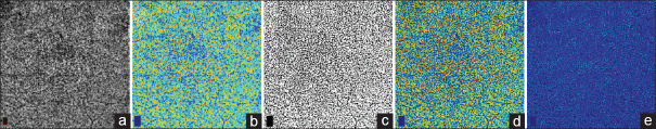
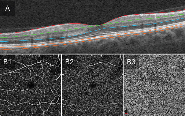
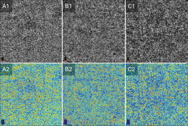
Materials and methods: In this retrospective study, 191 macular OCTA images were analyzed from 78 healthy eyes, 64 eyes from diabetic individuals without clinical signs of DR (NoDR), and 49 eyes with mild nonproliferative DR (NPDR). Five CC biomarkers were extracted from 6 mm × 6 mm enface OCTA images: flow deficit density (FDD), FD number (FDN), mean FD size (MFDS), perfusion intensity density (PID), and normalized blood flow index (NBFI). Flow maps were binarized using Phansalkar local thresholding, and statistical comparisons were performed using one-way analysis of variance and two-sample t-tests.
Results: All five biomarkers demonstrated significant differences across study groups (P < 0.001). FDD and MFDS were significantly elevated in both NoDR and mild NPDR eyes compared to controls, indicating increased nonperfusion and enlargement of flow voids. FDN decreased with disease severity, indicating spatial consolidation of capillary loss. PID and NBFI, which reflect flow signal intensity, also declined in diabetic eyes, suggesting a reduction in overall CC perfusion consistent with early vascular compromise.
Conclusion: Quantitative OCTA biomarkers of the CC reveal early microvascular changes in diabetic eyes. Among them, FDN and MFDS demonstrated the highest sensitivity to early disease progression. These findings support the use of CC-derived OCTA features as potential imaging biomarkers for detecting and monitoring early diabetic microvascular dysfunction.

 求助内容:
求助内容: 应助结果提醒方式:


