Kilohertz volumetric imaging of in vivo dynamics using squeezed light field microscopy
IF 32.1
1区 生物学
Q1 BIOCHEMICAL RESEARCH METHODS
引用次数: 0
Abstract
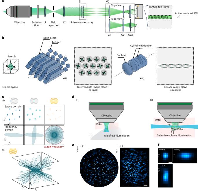
Volumetric functional imaging of transient cellular signaling and motion dynamics is often limited by hardware bandwidth and the scarcity of photons under short exposures. To overcome these challenges, we introduce squeezed light field microscopy (SLIM), a computational imaging approach that rapidly captures high-resolution three-dimensional light signals using only a single, low-format camera sensor. SLIM records over 1,000 volumes per second across a 550-µm diameter field of view and 300-µm depth, achieving 3.6-µm lateral and 6-µm axial resolution. Here we demonstrate its utility in blood cell velocimetry within the embryonic zebrafish brain and in freely moving tails undergoing high-frequency swings. Millisecond-scale temporal resolution further enables precise voltage imaging of neural membrane potentials in the leech ganglion and hippocampus of behaving mice. Together, these results establish SLIM as a versatile and robust tool for high-speed volumetric microscopy across diverse biological systems. Squeezed light field microscopy (SLIM) combines ideas from tomography and compressed sensing with light field microscopy to enable volumetric imaging at kilohertz rates, as demonstrated in blood flow imaging in zebrafish and voltage imaging in leeches and mice.
利用压缩光场显微镜对体内动力学进行千赫兹体积成像。
瞬态细胞信号和运动动力学的体积功能成像通常受到硬件带宽和短曝光下光子稀缺的限制。为了克服这些挑战,我们引入了压缩光场显微镜(SLIM),这是一种计算成像方法,仅使用单个低格式相机传感器即可快速捕获高分辨率三维光信号。SLIM在直径550微米、深度300微米的视场内每秒记录超过1000个卷,实现3.6微米的横向分辨率和6微米的轴向分辨率。在这里,我们展示了它在胚胎斑马鱼大脑内的血细胞速度测量和在经历高频摆动的自由运动尾巴中的应用。毫秒级的时间分辨率进一步实现了行为小鼠水蛭神经节和海马神经膜电位的精确电压成像。总之,这些结果建立SLIM作为一个多功能和强大的工具,高速体积显微镜在不同的生物系统。
本文章由计算机程序翻译,如有差异,请以英文原文为准。
求助全文
约1分钟内获得全文
求助全文
来源期刊
Nature Methods
生物-生化研究方法
CiteScore
58.70
自引率
1.70%
发文量
326
审稿时长
1 months
期刊介绍:
Nature Methods is a monthly journal that focuses on publishing innovative methods and substantial enhancements to fundamental life sciences research techniques. Geared towards a diverse, interdisciplinary readership of researchers in academia and industry engaged in laboratory work, the journal offers new tools for research and emphasizes the immediate practical significance of the featured work. It publishes primary research papers and reviews recent technical and methodological advancements, with a particular interest in primary methods papers relevant to the biological and biomedical sciences. This includes methods rooted in chemistry with practical applications for studying biological problems.
 求助内容:
求助内容: 应助结果提醒方式:


