{"title":"Actinomycosis of the Gallbladder in a Young Diabetic Woman with Acute Lithiasis Cholecystitis: A Case Report and Review of the Literature.","authors":"Mojgan Akbarzadeh-Jahromi, Seyed Mohammad Kazem Tadayyon, Sahar Asadi, Neda Soleimani, Sahand Mohammadzadeh, Hossein Afrakhteh","doi":"10.2147/IMCRJ.S544642","DOIUrl":null,"url":null,"abstract":"<p><strong>Introduction: </strong>Actinomycosis is a rare, chronic bacterial infection caused by the genus Actinomyces. The cervicofacial form is most common, while gallbladder involvement is exceptionally uncommon, with fewer than 50 cases reported in the literature.</p><p><strong>Case report: </strong>A 21-year-old diabetic woman presented with one day of nausea, vomiting, and persistent right upper quadrant abdominal pain. Ultrasonography and computed tomography scan revealed an enlarged gallbladder with biliary sludge and a single gallstone. A preoperative diagnosis of acute cholecystitis was made, and cholecystectomy was performed. Histopathological examination confirmed acute cholecystitis and demonstrated numerous filamentous, gram-positive bacteria, consistent with actinomycosis. Following 6 months of penicillin therapy, the patient remains well with no clinical or radiological evidence of recurrence.</p><p><strong>Conclusion: </strong>This case underscores the importance of routine histopathological evaluation of gallbladder specimens. Although rare, gallbladder actinomycosis should be considered in the differential diagnosis of gallbladder disease, particularly in immunocompromised individuals.</p>","PeriodicalId":14337,"journal":{"name":"International Medical Case Reports Journal","volume":"18 ","pages":"1225-1230"},"PeriodicalIF":0.7000,"publicationDate":"2025-09-18","publicationTypes":"Journal Article","fieldsOfStudy":null,"isOpenAccess":false,"openAccessPdf":"https://www.ncbi.nlm.nih.gov/pmc/articles/PMC12452965/pdf/","citationCount":"0","resultStr":null,"platform":"Semanticscholar","paperid":null,"PeriodicalName":"International Medical Case Reports Journal","FirstCategoryId":"1085","ListUrlMain":"https://doi.org/10.2147/IMCRJ.S544642","RegionNum":0,"RegionCategory":null,"ArticlePicture":[],"TitleCN":null,"AbstractTextCN":null,"PMCID":null,"EPubDate":"2025/1/1 0:00:00","PubModel":"eCollection","JCR":"Q3","JCRName":"MEDICINE, GENERAL & INTERNAL","Score":null,"Total":0}
引用次数: 0
Abstract
Introduction: Actinomycosis is a rare, chronic bacterial infection caused by the genus Actinomyces. The cervicofacial form is most common, while gallbladder involvement is exceptionally uncommon, with fewer than 50 cases reported in the literature.
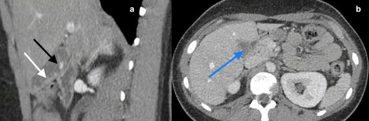
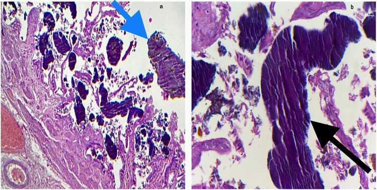
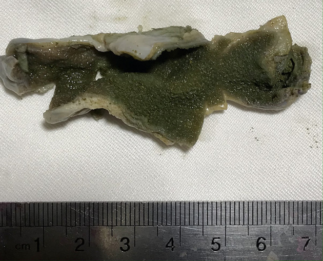
Case report: A 21-year-old diabetic woman presented with one day of nausea, vomiting, and persistent right upper quadrant abdominal pain. Ultrasonography and computed tomography scan revealed an enlarged gallbladder with biliary sludge and a single gallstone. A preoperative diagnosis of acute cholecystitis was made, and cholecystectomy was performed. Histopathological examination confirmed acute cholecystitis and demonstrated numerous filamentous, gram-positive bacteria, consistent with actinomycosis. Following 6 months of penicillin therapy, the patient remains well with no clinical or radiological evidence of recurrence.
Conclusion: This case underscores the importance of routine histopathological evaluation of gallbladder specimens. Although rare, gallbladder actinomycosis should be considered in the differential diagnosis of gallbladder disease, particularly in immunocompromised individuals.
期刊介绍:
International Medical Case Reports Journal is an international, peer-reviewed, open access, online journal publishing original case reports from all medical specialties. Submissions should not normally exceed 3,000 words or 4 published pages including figures, diagrams and references. As of 1st April 2019, the International Medical Case Reports Journal will no longer consider meta-analyses for publication.

 求助内容:
求助内容: 应助结果提醒方式:


