Deema Ammar Quzeih, Rasha A Alamoush, Sanaa Aljamani, Kifah Dafi Jamani
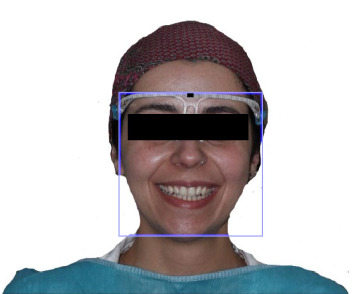
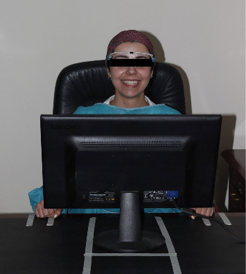
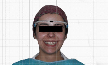
{"title":"Evaluating Canine Positioning Relative to Facial Landmarks: A Study on the Accuracy of the Inner Canthus and Alar of the Nose as Reference Points.","authors":"Deema Ammar Quzeih, Rasha A Alamoush, Sanaa Aljamani, Kifah Dafi Jamani","doi":"10.1155/ijod/8746060","DOIUrl":null,"url":null,"abstract":"<p><p><b>Background:</b> Accurately placing canine teeth in the dental arch is critical in achieving optimal esthetic and occlusal harmony, especially in edentulous patients. However, there is a lack of scientifically validated information to accurately place it in its true position. <b>Objective:</b> This study aimed to verify whether the inner canthus and alar of the nose reference points align exactly with the canine cusp tip position for both genders, providing valuable insights to enhance esthetic treatment outcomes. <b>Methods:</b> A total of 400 dental students aged between 20 and 30 years were enrolled in this study. A standardized frontal-view image of each participant was taken using a Lenovo computer camera that was set in a particular position throughout the process. The collected data was analyzed using Adobe Photoshop CSS software to determine the canine position, and the distance between the alar line and canine cusp tip line (ACT) was measured, as well as the distance from the inner canthus line to the canine cusp tip line (ICT) for both sides. Statistical analysis was performed using the independent <i>t</i>-test and chi-square test at a significance level of <i>p</i> < 0.05. <b>Results:</b> The alar of the nose line is located distally from the tip of the canine, indicating no precise alignment with the canine cusp tip, with a significant difference on both sides (<i>p</i> < 0.001). It was seen that the inner canthus line was located mesially, approximately 2 mm away from the canine cusp tip line, with no significant differences between genders (<i>p</i> > 0.05), and the same observation was seen for the right and left sides. <b>Conclusion:</b> The inner canthus of the eye and alar of the nose do not serve as a precise guide for positioning the canine cusp tip.</p>","PeriodicalId":13947,"journal":{"name":"International Journal of Dentistry","volume":"2025 ","pages":"8746060"},"PeriodicalIF":2.2000,"publicationDate":"2025-09-15","publicationTypes":"Journal Article","fieldsOfStudy":null,"isOpenAccess":false,"openAccessPdf":"https://www.ncbi.nlm.nih.gov/pmc/articles/PMC12453923/pdf/","citationCount":"0","resultStr":null,"platform":"Semanticscholar","paperid":null,"PeriodicalName":"International Journal of Dentistry","FirstCategoryId":"1085","ListUrlMain":"https://doi.org/10.1155/ijod/8746060","RegionNum":0,"RegionCategory":null,"ArticlePicture":[],"TitleCN":null,"AbstractTextCN":null,"PMCID":null,"EPubDate":"2025/1/1 0:00:00","PubModel":"eCollection","JCR":"Q2","JCRName":"DENTISTRY, ORAL SURGERY & MEDICINE","Score":null,"Total":0}
引用次数: 0
Abstract
Background: Accurately placing canine teeth in the dental arch is critical in achieving optimal esthetic and occlusal harmony, especially in edentulous patients. However, there is a lack of scientifically validated information to accurately place it in its true position. Objective: This study aimed to verify whether the inner canthus and alar of the nose reference points align exactly with the canine cusp tip position for both genders, providing valuable insights to enhance esthetic treatment outcomes. Methods: A total of 400 dental students aged between 20 and 30 years were enrolled in this study. A standardized frontal-view image of each participant was taken using a Lenovo computer camera that was set in a particular position throughout the process. The collected data was analyzed using Adobe Photoshop CSS software to determine the canine position, and the distance between the alar line and canine cusp tip line (ACT) was measured, as well as the distance from the inner canthus line to the canine cusp tip line (ICT) for both sides. Statistical analysis was performed using the independent t-test and chi-square test at a significance level of p < 0.05. Results: The alar of the nose line is located distally from the tip of the canine, indicating no precise alignment with the canine cusp tip, with a significant difference on both sides (p < 0.001). It was seen that the inner canthus line was located mesially, approximately 2 mm away from the canine cusp tip line, with no significant differences between genders (p > 0.05), and the same observation was seen for the right and left sides. Conclusion: The inner canthus of the eye and alar of the nose do not serve as a precise guide for positioning the canine cusp tip.

 求助内容:
求助内容: 应助结果提醒方式:


