Maria Kundzierewicz, Katarzyna Kołodziej, Arif Khokhar, Tsai Tsung-Ying, Artur Leśniak, Pawel Zakrzewski, Hubert Borecki, Ewelina Bohn, Jan Hecko, Jaroslav Januska, Daniel Precek, Maciej Stanuch, Andrzej Skalski, Yoshinobu Onuma, Patrick Serruys, Nico Bruining, Adriana Złahoda-Huzior, Dariusz Dudek
{"title":"Catheterization laboratories open the doors for Extended Realities-review of clinical applications in cardiology.","authors":"Maria Kundzierewicz, Katarzyna Kołodziej, Arif Khokhar, Tsai Tsung-Ying, Artur Leśniak, Pawel Zakrzewski, Hubert Borecki, Ewelina Bohn, Jan Hecko, Jaroslav Januska, Daniel Precek, Maciej Stanuch, Andrzej Skalski, Yoshinobu Onuma, Patrick Serruys, Nico Bruining, Adriana Złahoda-Huzior, Dariusz Dudek","doi":"10.1093/ehjdh/ztaf072","DOIUrl":null,"url":null,"abstract":"<p><p>The complexity and spatial relationships between vascular and cardiac structures, as well as anatomical diversity, pose a challenge for planning and performing cardiac interventions. Medical imaging, especially precise three-dimensional imaging techniques, plays a key role in the decision-making process. While traditional imaging methods like angiography, echocardiography, computed tomography, and magnetic resonance imaging remain gold standards, they have limitations in representing spatial relationships effectively. To overcome these limitations, advanced techniques such as three-dimensional printing, three-dimensional modelling, and Extended Realities are needed. Focusing on Extended Realities, their main advantages are direct spatial visualization based on medical data, interaction with objects, and immersion in cardiac anatomy. These benefits impact procedural planning and intra-procedural navigation. The following publication presents current applications, benefits, drawbacks, and limitations of Virtual, Augmented, and Mixed Reality technologies in cardiac interventions. The aim of this review is to improve understanding and utilization of the entire spectrum of these innovative tools in clinical practice.</p>","PeriodicalId":72965,"journal":{"name":"European heart journal. Digital health","volume":"6 5","pages":"1055-1068"},"PeriodicalIF":4.4000,"publicationDate":"2025-06-23","publicationTypes":"Journal Article","fieldsOfStudy":null,"isOpenAccess":false,"openAccessPdf":"https://www.ncbi.nlm.nih.gov/pmc/articles/PMC12450514/pdf/","citationCount":"0","resultStr":null,"platform":"Semanticscholar","paperid":null,"PeriodicalName":"European heart journal. Digital health","FirstCategoryId":"1085","ListUrlMain":"https://doi.org/10.1093/ehjdh/ztaf072","RegionNum":0,"RegionCategory":null,"ArticlePicture":[],"TitleCN":null,"AbstractTextCN":null,"PMCID":null,"EPubDate":"2025/9/1 0:00:00","PubModel":"eCollection","JCR":"Q1","JCRName":"CARDIAC & CARDIOVASCULAR SYSTEMS","Score":null,"Total":0}
引用次数: 0
Abstract
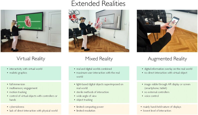
The complexity and spatial relationships between vascular and cardiac structures, as well as anatomical diversity, pose a challenge for planning and performing cardiac interventions. Medical imaging, especially precise three-dimensional imaging techniques, plays a key role in the decision-making process. While traditional imaging methods like angiography, echocardiography, computed tomography, and magnetic resonance imaging remain gold standards, they have limitations in representing spatial relationships effectively. To overcome these limitations, advanced techniques such as three-dimensional printing, three-dimensional modelling, and Extended Realities are needed. Focusing on Extended Realities, their main advantages are direct spatial visualization based on medical data, interaction with objects, and immersion in cardiac anatomy. These benefits impact procedural planning and intra-procedural navigation. The following publication presents current applications, benefits, drawbacks, and limitations of Virtual, Augmented, and Mixed Reality technologies in cardiac interventions. The aim of this review is to improve understanding and utilization of the entire spectrum of these innovative tools in clinical practice.


 求助内容:
求助内容: 应助结果提醒方式:


