{"title":"Artificial intelligence-based accurate myocardial infarction mapping using 12-lead electrocardiography.","authors":"Hui Wang, Zhifan Gao, Heye Zhang, Yuzhen Zhu, Shichang Lian, Kairui Bo, Shuang Li, Yifeng Gao, Baiyan Zhuang, Zhen Zhou, Xinwei Zhang, Cuiyan Wang, Koen Nieman, Lei Xu","doi":"10.1093/ehjdh/ztaf077","DOIUrl":null,"url":null,"abstract":"<p><strong>Aims: </strong>Assessing myocardial fibrosis (MF) in patients with prior myocardial infarction (MI) is crucial for prognosis. Artificial intelligence-assisted electrocardiography (AI-ECG) has a great potential to detect MF. However, training a precise AI-ECG model requires voluminous ECGs. A biosimulation model may be an efficient substitution. This study aimed to develop and validate a novel artificial intelligence-assisted method using 12-lead electrocardiography (AI-MI-12ECG).</p><p><strong>Methods and results: </strong>The AI-MI-12ECG was trained by a biosimulation model to visualize the presence, location, and size of MF in post-MI patients. A total of 182 post-MI patients were included in this prospective study. The MF detected by AI-MI-12ECG and the cardiologist were compared with the late gadolinium-enhanced (LGE) area of cardiac magnetic resonance (CMR). The results show that AI-MI-12ECG exhibited strong correlation with LGE in identifying the MI location (<i>R</i> = 0.955). Compared with CMR-LGE, AI-MI-12ECG achieved receiver operating characteristic curves of 0.95, 0.95, and 0.89 for left anterior descending coronary artery (LAD), right coronary artery (RCA), and left circumflex coronary artery (LCX) territories, respectively, with high accuracies for LAD (0.95), RCA (0.97), and LCX (0.91).</p><p><strong>Conclusion: </strong>The AI-MI-12ECG trained using the biosimulation model in post-MI patients was adequately aligned with CMR-LGE. This highlights its potential for accurate detection of fibrosis and identification of individuals with significant infarct burdens.</p>","PeriodicalId":72965,"journal":{"name":"European heart journal. Digital health","volume":"6 5","pages":"939-948"},"PeriodicalIF":4.4000,"publicationDate":"2025-07-01","publicationTypes":"Journal Article","fieldsOfStudy":null,"isOpenAccess":false,"openAccessPdf":"https://www.ncbi.nlm.nih.gov/pmc/articles/PMC12450511/pdf/","citationCount":"0","resultStr":null,"platform":"Semanticscholar","paperid":null,"PeriodicalName":"European heart journal. Digital health","FirstCategoryId":"1085","ListUrlMain":"https://doi.org/10.1093/ehjdh/ztaf077","RegionNum":0,"RegionCategory":null,"ArticlePicture":[],"TitleCN":null,"AbstractTextCN":null,"PMCID":null,"EPubDate":"2025/9/1 0:00:00","PubModel":"eCollection","JCR":"Q1","JCRName":"CARDIAC & CARDIOVASCULAR SYSTEMS","Score":null,"Total":0}
引用次数: 0
Abstract
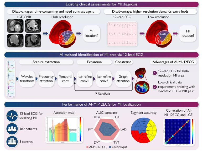
Aims: Assessing myocardial fibrosis (MF) in patients with prior myocardial infarction (MI) is crucial for prognosis. Artificial intelligence-assisted electrocardiography (AI-ECG) has a great potential to detect MF. However, training a precise AI-ECG model requires voluminous ECGs. A biosimulation model may be an efficient substitution. This study aimed to develop and validate a novel artificial intelligence-assisted method using 12-lead electrocardiography (AI-MI-12ECG).
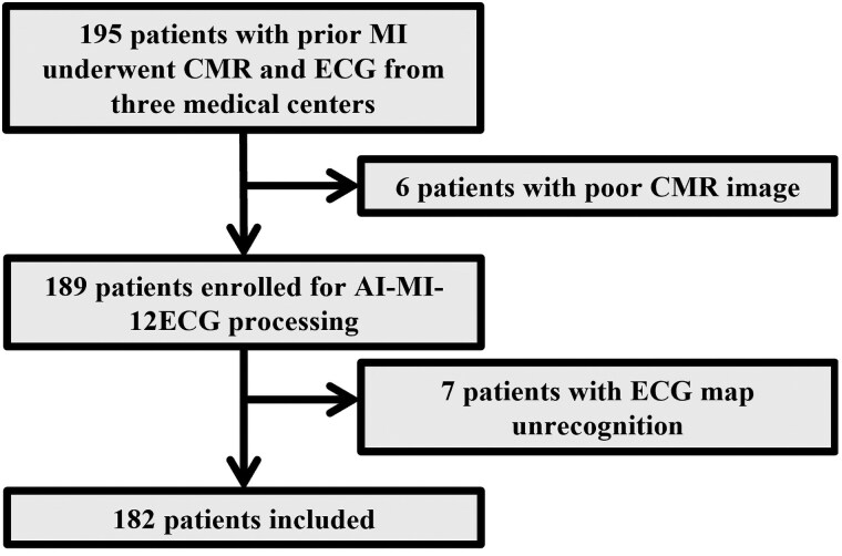
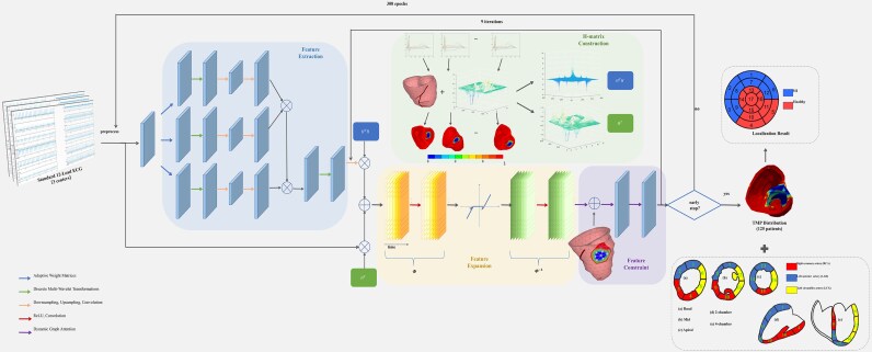
Methods and results: The AI-MI-12ECG was trained by a biosimulation model to visualize the presence, location, and size of MF in post-MI patients. A total of 182 post-MI patients were included in this prospective study. The MF detected by AI-MI-12ECG and the cardiologist were compared with the late gadolinium-enhanced (LGE) area of cardiac magnetic resonance (CMR). The results show that AI-MI-12ECG exhibited strong correlation with LGE in identifying the MI location (R = 0.955). Compared with CMR-LGE, AI-MI-12ECG achieved receiver operating characteristic curves of 0.95, 0.95, and 0.89 for left anterior descending coronary artery (LAD), right coronary artery (RCA), and left circumflex coronary artery (LCX) territories, respectively, with high accuracies for LAD (0.95), RCA (0.97), and LCX (0.91).
Conclusion: The AI-MI-12ECG trained using the biosimulation model in post-MI patients was adequately aligned with CMR-LGE. This highlights its potential for accurate detection of fibrosis and identification of individuals with significant infarct burdens.

 求助内容:
求助内容: 应助结果提醒方式:


