Mikaela Engarås Hamre, Lise Benedikte Wendt Ræder, Martin Okelsrud Riiser, Peter Franz Schubert, Marius Molund
{"title":"Metatarsal Pronation on Radiographs: A Prospective Reliability Study of Visual Rotation Markers in Hallux Valgus.","authors":"Mikaela Engarås Hamre, Lise Benedikte Wendt Ræder, Martin Okelsrud Riiser, Peter Franz Schubert, Marius Molund","doi":"10.1177/24730114251371723","DOIUrl":null,"url":null,"abstract":"<p><strong>Background: </strong>Rotational malalignment of the first metatarsal is increasingly recognized as a key feature of hallux valgus deformity, but the reliability of radiographic rotation markers remains uncertain. This study assessed the inter- and intraobserver reliability of 4 commonly used radiographic parameters: metatarsal pronation angle (MPA), tibial sesamoid position (TSP), lateral head shape (LHS), and round head sign (RH).</p><p><strong>Methods: </strong>In this prospective reliability study, 3 senior clinicians independently evaluated weightbearing anteroposterior and axial sesamoid radiographs of 75 hallux valgus cases on 2 occasions. Metatarsal pronation angle (MPA) was measured as a continuous variable and analyzed using intraclass correlation coefficients (ICCs). Tibial sesamoid position (TSP), lateral head shape (LHS), and round head sign (RH) were graded using ordinal scales and assessed with weighted kappa statistics (κ). Subgroup analyses evaluated whether reliability varied by deformity severity (hallux valgus angle) or increased distal metatarsal articular angle (DMAA > 10 degrees).</p><p><strong>Results: </strong>MPA showed excellent agreement (ICC = 0.81-0.94). TSP also demonstrated high reliability (κ = 0.88-0.98), although its value as a rotation marker is limited. LHS showed moderate to substantial agreement (κ = 0.59-0.85), whereas RH had fair to moderate reliability (κ = 0.35-0.66). RH was least reliable in mild deformities, whereas other parameters remained stable across subgroups, with slightly lower values in cases with elevated DMAA.</p><p><strong>Conclusions: </strong>Conventional radiographs offer reliable assessment of MPA and TSP. LHS provides acceptable reproducibility, whereas RH is less consistent. These findings support the use of selected radiographic markers and suggest that further validation against 3-dimensional imaging and standardized grading frameworks may improve consistency and clinical applicability.</p><p><strong>Level of evidence: </strong>Level IV, case series.</p>","PeriodicalId":12429,"journal":{"name":"Foot & Ankle Orthopaedics","volume":"10 3","pages":"24730114251371723"},"PeriodicalIF":0.0000,"publicationDate":"2025-09-20","publicationTypes":"Journal Article","fieldsOfStudy":null,"isOpenAccess":false,"openAccessPdf":"https://www.ncbi.nlm.nih.gov/pmc/articles/PMC12450261/pdf/","citationCount":"0","resultStr":null,"platform":"Semanticscholar","paperid":null,"PeriodicalName":"Foot & Ankle Orthopaedics","FirstCategoryId":"1085","ListUrlMain":"https://doi.org/10.1177/24730114251371723","RegionNum":0,"RegionCategory":null,"ArticlePicture":[],"TitleCN":null,"AbstractTextCN":null,"PMCID":null,"EPubDate":"2025/7/1 0:00:00","PubModel":"eCollection","JCR":"","JCRName":"","Score":null,"Total":0}
引用次数: 0
Abstract
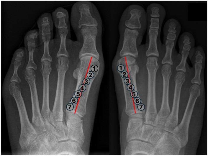
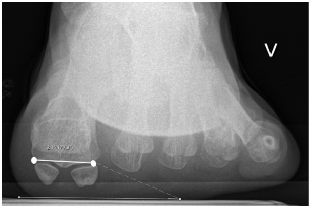
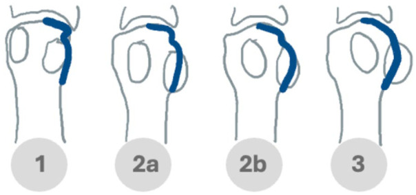
Background: Rotational malalignment of the first metatarsal is increasingly recognized as a key feature of hallux valgus deformity, but the reliability of radiographic rotation markers remains uncertain. This study assessed the inter- and intraobserver reliability of 4 commonly used radiographic parameters: metatarsal pronation angle (MPA), tibial sesamoid position (TSP), lateral head shape (LHS), and round head sign (RH).
Methods: In this prospective reliability study, 3 senior clinicians independently evaluated weightbearing anteroposterior and axial sesamoid radiographs of 75 hallux valgus cases on 2 occasions. Metatarsal pronation angle (MPA) was measured as a continuous variable and analyzed using intraclass correlation coefficients (ICCs). Tibial sesamoid position (TSP), lateral head shape (LHS), and round head sign (RH) were graded using ordinal scales and assessed with weighted kappa statistics (κ). Subgroup analyses evaluated whether reliability varied by deformity severity (hallux valgus angle) or increased distal metatarsal articular angle (DMAA > 10 degrees).
Results: MPA showed excellent agreement (ICC = 0.81-0.94). TSP also demonstrated high reliability (κ = 0.88-0.98), although its value as a rotation marker is limited. LHS showed moderate to substantial agreement (κ = 0.59-0.85), whereas RH had fair to moderate reliability (κ = 0.35-0.66). RH was least reliable in mild deformities, whereas other parameters remained stable across subgroups, with slightly lower values in cases with elevated DMAA.
Conclusions: Conventional radiographs offer reliable assessment of MPA and TSP. LHS provides acceptable reproducibility, whereas RH is less consistent. These findings support the use of selected radiographic markers and suggest that further validation against 3-dimensional imaging and standardized grading frameworks may improve consistency and clinical applicability.

 求助内容:
求助内容: 应助结果提醒方式:


