{"title":"Gray matter volume heterogeneity by stage, site of origin and pathophysiology in schizophrenia","authors":"Yuchao Jiang, Lena Palaniyappan, Xiao Chang, Jie Zhang, Enpeng Zhou, Xin Yu, Shih-Jen Tsai, Ching-Po Lin, Jingliang Cheng, Yingying Tang, Jijun Wang, Cheng Luo, Dezhong Yao, Long-Biao Cui, Wei Cheng, Jianfeng Feng","doi":"10.1038/s44220-025-00449-9","DOIUrl":null,"url":null,"abstract":"Schizophrenia is characterized with greater variability beyond the mean differences in brain structures. This variability is assumed to be static, reflecting the presence of heterogeneous subgroups, but this assumption and alternative explanations remain untested. Here we examine whether gray matter volume variability decreases in later stages of schizophrenia using magnetic resonance imaging of 1,792 individuals with schizophrenia and 1,523 healthy controls. Compared with healthy controls, greater variability (false-discovery-rate-corrected P < 0.05) was found in 50 regions across the entire patient group. The average variability across all regions was greater in the first-episode than chronic stage (t = 10.8, P = 1.7 × 10⁻⁷). The areas with the largest variability were located at the frontotemporal cortex and thalamus (first-episode), or the hippocampus and caudate (chronic). This study offers novel insights into the diversity of brain alterations in schizophrenia, emphasizing that brain-based heterogeneity is not a static feature; it is more pronounced at the onset of the disorder but reduced over the long term. Schizophrenia exhibits significant variability in brain structures, traditionally viewed as static due to heterogeneous subgroups. Here the authors use magnetic resonance imaging data from 1,792 individuals with schizophrenia to reveal that gray matter volume variability is greater in early stages and decreases over time, highlighting dynamic brain alterations with implications for understanding disease progression.","PeriodicalId":74247,"journal":{"name":"Nature mental health","volume":"3 7","pages":"803-813"},"PeriodicalIF":8.7000,"publicationDate":"2025-06-23","publicationTypes":"Journal Article","fieldsOfStudy":null,"isOpenAccess":false,"openAccessPdf":"","citationCount":"0","resultStr":null,"platform":"Semanticscholar","paperid":null,"PeriodicalName":"Nature mental health","FirstCategoryId":"1085","ListUrlMain":"https://www.nature.com/articles/s44220-025-00449-9","RegionNum":0,"RegionCategory":null,"ArticlePicture":[],"TitleCN":null,"AbstractTextCN":null,"PMCID":null,"EPubDate":"","PubModel":"","JCR":"","JCRName":"","Score":null,"Total":0}
引用次数: 0
Abstract
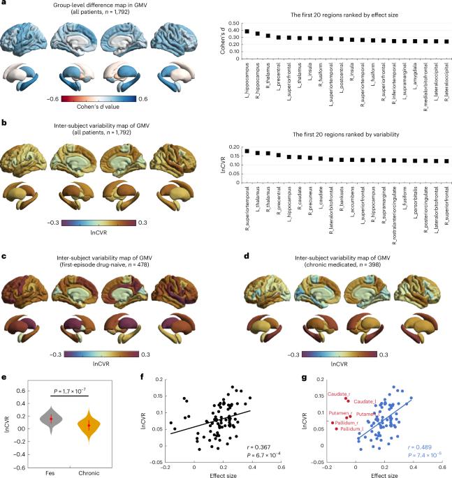
Schizophrenia is characterized with greater variability beyond the mean differences in brain structures. This variability is assumed to be static, reflecting the presence of heterogeneous subgroups, but this assumption and alternative explanations remain untested. Here we examine whether gray matter volume variability decreases in later stages of schizophrenia using magnetic resonance imaging of 1,792 individuals with schizophrenia and 1,523 healthy controls. Compared with healthy controls, greater variability (false-discovery-rate-corrected P < 0.05) was found in 50 regions across the entire patient group. The average variability across all regions was greater in the first-episode than chronic stage (t = 10.8, P = 1.7 × 10⁻⁷). The areas with the largest variability were located at the frontotemporal cortex and thalamus (first-episode), or the hippocampus and caudate (chronic). This study offers novel insights into the diversity of brain alterations in schizophrenia, emphasizing that brain-based heterogeneity is not a static feature; it is more pronounced at the onset of the disorder but reduced over the long term. Schizophrenia exhibits significant variability in brain structures, traditionally viewed as static due to heterogeneous subgroups. Here the authors use magnetic resonance imaging data from 1,792 individuals with schizophrenia to reveal that gray matter volume variability is greater in early stages and decreases over time, highlighting dynamic brain alterations with implications for understanding disease progression.

 求助内容:
求助内容: 应助结果提醒方式:


