Xiuyun Lu, Jianqing Ye, Xiuwu Pan, Shaojun Chen, Liang Zhang, Ying Wang, Juan Cheng, Jiaying Cao, Li Wei, Xingang Cui, Yi Dong
{"title":"Analysis of correlation between dynamic contrast-enhanced ultrasound and angiogenesis activity of renal cell carcinoma.","authors":"Xiuyun Lu, Jianqing Ye, Xiuwu Pan, Shaojun Chen, Liang Zhang, Ying Wang, Juan Cheng, Jiaying Cao, Li Wei, Xingang Cui, Yi Dong","doi":"10.1055/a-2513-1054","DOIUrl":null,"url":null,"abstract":"<p><strong>Purpose: </strong>To investigate the potential correlation of dynamic contrast-enhanced ultrasound (DCE-US) with angiogenesis activity of renal cell carcinoma (RCC).</p><p><strong>Materials and methods: </strong>Patients with surgery resection and histopathologically proven RCC lesions were included. B-mode ultrasound (BMUS) and contrast-enhanced ultrasound (CEUS) were performed one week before surgery. SonoVue was injected as the contrast agent. VueBox (Bracco, Italy) was used for the quantitative analysis. According to the histopathological and immunohistochemical results, patients were classified into two groups: active angiogenesis and inactive angiogenesis. Time intensity curves (TICs) and quantitative parameters were compared between two groups.</p><p><strong>Results: </strong>From July 2023 to November 2023, a total of 50 patients (13 females and 37 males, mean age 61.1±11.1 years) were included. The mean size of the lesions was 39.4±2.7 mm. Patients were classified into the active angiogenesis group (n=30) and the inactive angiogenesis group (n=20). On BMUS, 68.0% (34/50) of RCCs were visualized as hypoechoic lesions with ill-defined borders and irregular shapes (P>0.05). During cortical phase of CEUS, 72.6% (23/30) of RCCs with active angiogenesis were visualized with hyperenhancement (P=0.027). Only 30.0% (9/30) of RCCs with active angiogenesis showed hypo-enhancement in the parenchymal phase (P>0.05). Compared to the inactive angiogenesis group, TICs of the active angiogenesis group revealed faster and greater enhancement in the cortical phase, slower decline during the parenchymal phase, and an increased area under the curve. Among quantitative parameters, the active angiogenesis group showed the higher ratio of wash-in rate and wash-in perfusion index (P<0.05).</p><p><strong>Conclusion: </strong>DCE-US analysis has potential value in predicting angiogenesis activity in RCC lesions.</p>","PeriodicalId":44852,"journal":{"name":"Ultrasound International Open","volume":"11 ","pages":"a25131054"},"PeriodicalIF":1.6000,"publicationDate":"2025-09-15","publicationTypes":"Journal Article","fieldsOfStudy":null,"isOpenAccess":false,"openAccessPdf":"https://www.ncbi.nlm.nih.gov/pmc/articles/PMC12445333/pdf/","citationCount":"0","resultStr":null,"platform":"Semanticscholar","paperid":null,"PeriodicalName":"Ultrasound International Open","FirstCategoryId":"1085","ListUrlMain":"https://doi.org/10.1055/a-2513-1054","RegionNum":0,"RegionCategory":null,"ArticlePicture":[],"TitleCN":null,"AbstractTextCN":null,"PMCID":null,"EPubDate":"2025/1/1 0:00:00","PubModel":"eCollection","JCR":"Q3","JCRName":"RADIOLOGY, NUCLEAR MEDICINE & MEDICAL IMAGING","Score":null,"Total":0}
引用次数: 0
Abstract
Purpose: To investigate the potential correlation of dynamic contrast-enhanced ultrasound (DCE-US) with angiogenesis activity of renal cell carcinoma (RCC).
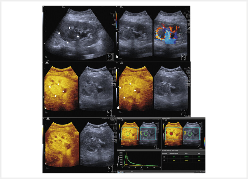
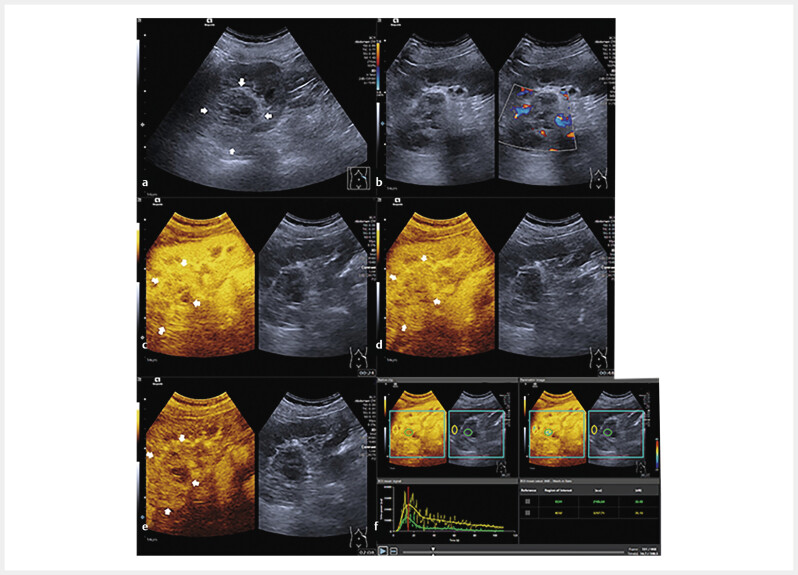
Materials and methods: Patients with surgery resection and histopathologically proven RCC lesions were included. B-mode ultrasound (BMUS) and contrast-enhanced ultrasound (CEUS) were performed one week before surgery. SonoVue was injected as the contrast agent. VueBox (Bracco, Italy) was used for the quantitative analysis. According to the histopathological and immunohistochemical results, patients were classified into two groups: active angiogenesis and inactive angiogenesis. Time intensity curves (TICs) and quantitative parameters were compared between two groups.
Results: From July 2023 to November 2023, a total of 50 patients (13 females and 37 males, mean age 61.1±11.1 years) were included. The mean size of the lesions was 39.4±2.7 mm. Patients were classified into the active angiogenesis group (n=30) and the inactive angiogenesis group (n=20). On BMUS, 68.0% (34/50) of RCCs were visualized as hypoechoic lesions with ill-defined borders and irregular shapes (P>0.05). During cortical phase of CEUS, 72.6% (23/30) of RCCs with active angiogenesis were visualized with hyperenhancement (P=0.027). Only 30.0% (9/30) of RCCs with active angiogenesis showed hypo-enhancement in the parenchymal phase (P>0.05). Compared to the inactive angiogenesis group, TICs of the active angiogenesis group revealed faster and greater enhancement in the cortical phase, slower decline during the parenchymal phase, and an increased area under the curve. Among quantitative parameters, the active angiogenesis group showed the higher ratio of wash-in rate and wash-in perfusion index (P<0.05).
Conclusion: DCE-US analysis has potential value in predicting angiogenesis activity in RCC lesions.

 求助内容:
求助内容: 应助结果提醒方式:


