Evaluation of maturation-related changes in maxillary sinus diameter and cheek teeth positioning relative to the maxillary sinus in the Dareshuri horse.
{"title":"Evaluation of maturation-related changes in maxillary sinus diameter and cheek teeth positioning relative to the maxillary sinus in the Dareshuri horse.","authors":"Zahra Riahi, Aboutorab Tabatabaei Naeini, Reza Zare","doi":"10.1294/jes.36.81","DOIUrl":null,"url":null,"abstract":"<p><p>Dareshuri horses are the predominant breed in Fars Province, Iran. Although disorders affecting their maxillary cheek teeth and maxillary sinuses are relatively common, limited fundamental data are available on the dimensions and relationships of these structures at different ages. Given the significant anatomical changes in the heads of young horses as they mature, this study aimed to evaluate age-related changes in the position and anatomical relationships of individual maxillary cheek teeth within the rostral and caudal maxillary sinuses (RMS and CMS, respectively), as well as changes in the lengths and heights of individual sinus compartments during their growth. Radiographs were performed on 29 heads of live, healthy horses aged between 4 months and 5 years and were analyzed using the EConsole1 Radiography Viewer software (V.3, 2017, DRTECH Europe GmbH, Schwalbach am Taunus, Germany). Statistical analyses revealed that the only significant change throughout the study was an increase in the length of the CMS (4.075 ± 0.99 cm; SE), which was more significant in horses up to three years old. At less than 1 year old, the only tooth present in the maxillary sinus was M1. At 1-2 years old, M2 was observed entering the maxillary compartments; PM4 entered the RMS at 2-3 years old, and M3 entered the CMS at 3-4 years old. Eventually at 4-5 years old, PM3, M1, and M2 were present in the RMS, and M2 and M3 were present in the CMS. This information should be of value in the diagnosis and treatment of Dareshuri maxillofacial disorders and used as a reference for further anatomical investigations.</p>","PeriodicalId":35701,"journal":{"name":"Journal of Equine Science","volume":"36 3","pages":"81-91"},"PeriodicalIF":0.0000,"publicationDate":"2025-01-01","publicationTypes":"Journal Article","fieldsOfStudy":null,"isOpenAccess":false,"openAccessPdf":"https://www.ncbi.nlm.nih.gov/pmc/articles/PMC12445997/pdf/","citationCount":"0","resultStr":null,"platform":"Semanticscholar","paperid":null,"PeriodicalName":"Journal of Equine Science","FirstCategoryId":"1085","ListUrlMain":"https://doi.org/10.1294/jes.36.81","RegionNum":0,"RegionCategory":null,"ArticlePicture":[],"TitleCN":null,"AbstractTextCN":null,"PMCID":null,"EPubDate":"2025/9/17 0:00:00","PubModel":"Epub","JCR":"Q3","JCRName":"Veterinary","Score":null,"Total":0}
引用次数: 0
Abstract
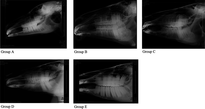
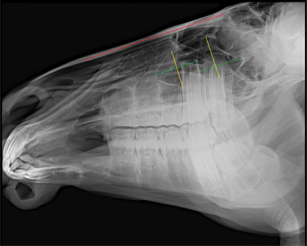
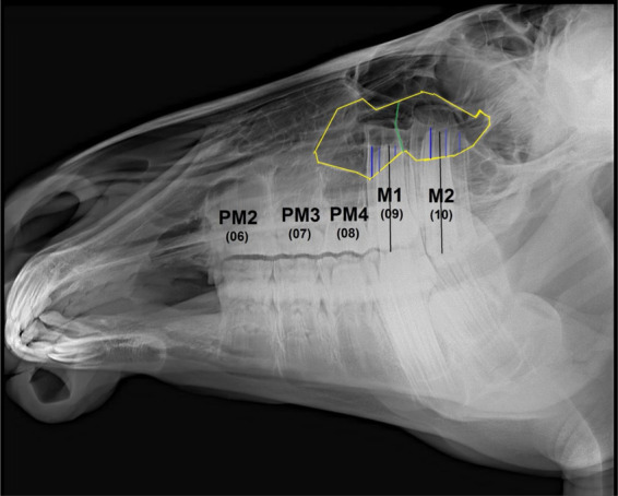
Dareshuri horses are the predominant breed in Fars Province, Iran. Although disorders affecting their maxillary cheek teeth and maxillary sinuses are relatively common, limited fundamental data are available on the dimensions and relationships of these structures at different ages. Given the significant anatomical changes in the heads of young horses as they mature, this study aimed to evaluate age-related changes in the position and anatomical relationships of individual maxillary cheek teeth within the rostral and caudal maxillary sinuses (RMS and CMS, respectively), as well as changes in the lengths and heights of individual sinus compartments during their growth. Radiographs were performed on 29 heads of live, healthy horses aged between 4 months and 5 years and were analyzed using the EConsole1 Radiography Viewer software (V.3, 2017, DRTECH Europe GmbH, Schwalbach am Taunus, Germany). Statistical analyses revealed that the only significant change throughout the study was an increase in the length of the CMS (4.075 ± 0.99 cm; SE), which was more significant in horses up to three years old. At less than 1 year old, the only tooth present in the maxillary sinus was M1. At 1-2 years old, M2 was observed entering the maxillary compartments; PM4 entered the RMS at 2-3 years old, and M3 entered the CMS at 3-4 years old. Eventually at 4-5 years old, PM3, M1, and M2 were present in the RMS, and M2 and M3 were present in the CMS. This information should be of value in the diagnosis and treatment of Dareshuri maxillofacial disorders and used as a reference for further anatomical investigations.

 求助内容:
求助内容: 应助结果提醒方式:


