Fabrication of 3D Collagen-Based Decellularized Biological Scaffolds Using Human Wharton's Jelly-Derived Mesenchymal Stem Cells With Differentiation Potential Toward Chondrocytes.
{"title":"Fabrication of 3D Collagen-Based Decellularized Biological Scaffolds Using Human Wharton's Jelly-Derived Mesenchymal Stem Cells With Differentiation Potential Toward Chondrocytes.","authors":"Fatemeh Masjedi, Zahra Heidari, Kamran Hosseini, Shahrokh Zare, Anahid Safari, Davood Mehrabani, Elmira Jalilian, Negar Azarpira, Zahra Khodabandeh","doi":"10.1155/sci/9953810","DOIUrl":null,"url":null,"abstract":"<p><p><b>Background:</b> Stem cell-based regenerative approaches have been developed to treat osteoarthritis (OA) and repair cartilage defects. In the present study, we fabricated a three-dimensional (3D) collagen-based decellularized biological scaffold using human Wharton's jelly-derived mesenchymal stem cells (hWJ-MSCs) and analyzed its recellularization and subsequent differentiation potential toward chondrocytes. <b>Methods:</b> MSCs were isolated from human Wharton's jelly, characterized by flow cytometry, and differentiated toward osteogenic and adipogenic lineages. hWJ-MSCs were cultured in a 3D collagen scaffold. After the matrix was deposited by the cells, the scaffold was decellularized, and new hWJ-MSCs were cultured and differentiated into chondrocytes. The efficiency of the decellularization process was assessed using hematoxylin and eosin (H&E) staining, DNA quantification, scanning electron microscopy (SEM), and Raman spectroscopy. Immunohistochemical and transcriptional evaluation of chondrogenic markers, including collagen type II, aggrecan, and osteonectin, was performed. <b>Results:</b> Prepared decellularized scaffolds showed very low levels of nucleic materials compared to intact ones. The integrity and efficiency of the decellularization process were confirmed using SEM. Moreover, a comparison of Raman spectra of intact and decellularized scaffolds demonstrated a remarkable reduction in carbohydrate, lipid, and DNA content. Three weeks after recellularization in the presence of chondrogenic medium, the immunoreactivity and expression levels of specific chondrocyte markers, including collagen type II, aggrecan, and osteonectin, significantly increased compared to negative controls. <b>Conclusion:</b> hWJ-MSCs and their use in fabricating nucleic acid-free 3D collagen-based scaffolds represent a promising in vitro model for investigating how the extracellular matrix (ECM) contributes to specific cell microenvironments. Decellularized ECM can also be utilized to develop novel, cell-free biomedical products for regenerative medicine.</p>","PeriodicalId":21962,"journal":{"name":"Stem Cells International","volume":"2025 ","pages":"9953810"},"PeriodicalIF":3.3000,"publicationDate":"2025-09-11","publicationTypes":"Journal Article","fieldsOfStudy":null,"isOpenAccess":false,"openAccessPdf":"https://www.ncbi.nlm.nih.gov/pmc/articles/PMC12446596/pdf/","citationCount":"0","resultStr":null,"platform":"Semanticscholar","paperid":null,"PeriodicalName":"Stem Cells International","FirstCategoryId":"3","ListUrlMain":"https://doi.org/10.1155/sci/9953810","RegionNum":3,"RegionCategory":"医学","ArticlePicture":[],"TitleCN":null,"AbstractTextCN":null,"PMCID":null,"EPubDate":"2025/1/1 0:00:00","PubModel":"eCollection","JCR":"Q2","JCRName":"CELL & TISSUE ENGINEERING","Score":null,"Total":0}
引用次数: 0
Abstract
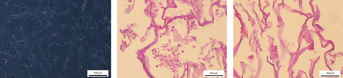
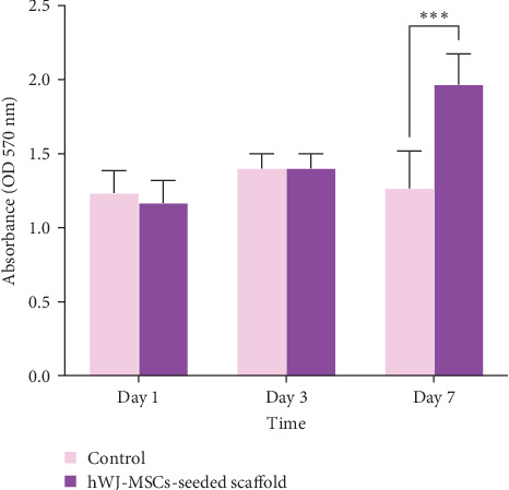
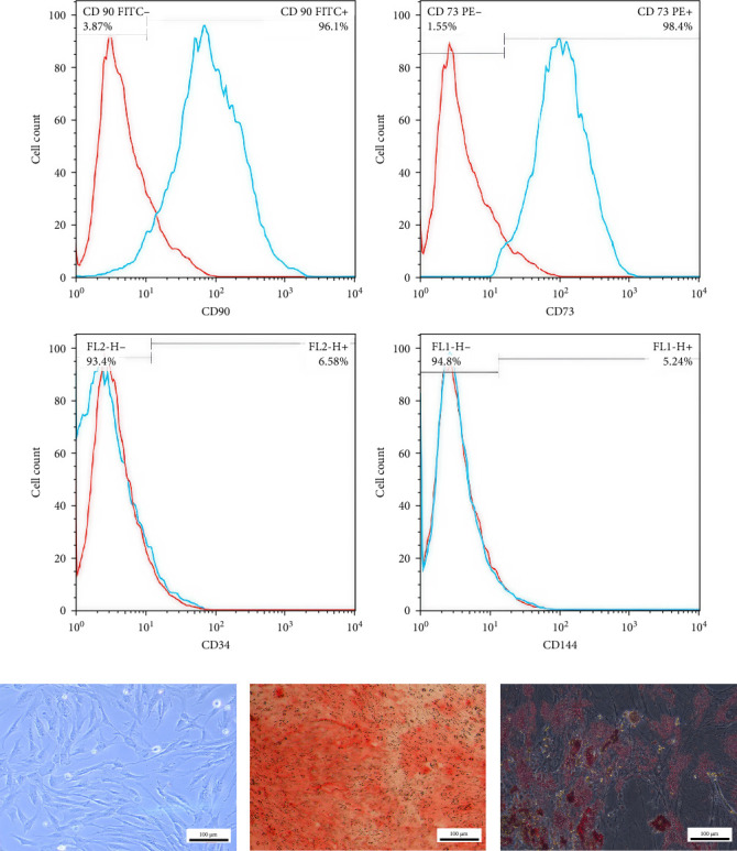
Background: Stem cell-based regenerative approaches have been developed to treat osteoarthritis (OA) and repair cartilage defects. In the present study, we fabricated a three-dimensional (3D) collagen-based decellularized biological scaffold using human Wharton's jelly-derived mesenchymal stem cells (hWJ-MSCs) and analyzed its recellularization and subsequent differentiation potential toward chondrocytes. Methods: MSCs were isolated from human Wharton's jelly, characterized by flow cytometry, and differentiated toward osteogenic and adipogenic lineages. hWJ-MSCs were cultured in a 3D collagen scaffold. After the matrix was deposited by the cells, the scaffold was decellularized, and new hWJ-MSCs were cultured and differentiated into chondrocytes. The efficiency of the decellularization process was assessed using hematoxylin and eosin (H&E) staining, DNA quantification, scanning electron microscopy (SEM), and Raman spectroscopy. Immunohistochemical and transcriptional evaluation of chondrogenic markers, including collagen type II, aggrecan, and osteonectin, was performed. Results: Prepared decellularized scaffolds showed very low levels of nucleic materials compared to intact ones. The integrity and efficiency of the decellularization process were confirmed using SEM. Moreover, a comparison of Raman spectra of intact and decellularized scaffolds demonstrated a remarkable reduction in carbohydrate, lipid, and DNA content. Three weeks after recellularization in the presence of chondrogenic medium, the immunoreactivity and expression levels of specific chondrocyte markers, including collagen type II, aggrecan, and osteonectin, significantly increased compared to negative controls. Conclusion: hWJ-MSCs and their use in fabricating nucleic acid-free 3D collagen-based scaffolds represent a promising in vitro model for investigating how the extracellular matrix (ECM) contributes to specific cell microenvironments. Decellularized ECM can also be utilized to develop novel, cell-free biomedical products for regenerative medicine.
期刊介绍:
Stem Cells International is a peer-reviewed, Open Access journal that publishes original research articles, review articles, and clinical studies in all areas of stem cell biology and applications. The journal will consider basic, translational, and clinical research, including animal models and clinical trials.
Topics covered include, but are not limited to: embryonic stem cells; induced pluripotent stem cells; tissue-specific stem cells; stem cell differentiation; genetics and epigenetics; cancer stem cells; stem cell technologies; ethical, legal, and social issues.
 求助内容:
求助内容: 应助结果提醒方式:


