Dilip Shettigar, Rajagopal Kadavigere, K Vaishali, Nitika C Panakkal, Winniecia Dkhar, Baskaran Chandrasekaran, Hari Prakash Palaniswamy, Sneha Ravichandran, Sathya Sabina Muthu, Poovitha Shruthi Paramashiva, Suresh Sukumar
{"title":"Impact of physical activity and body mass index on brain structure: A cross-sectional MRI study.","authors":"Dilip Shettigar, Rajagopal Kadavigere, K Vaishali, Nitika C Panakkal, Winniecia Dkhar, Baskaran Chandrasekaran, Hari Prakash Palaniswamy, Sneha Ravichandran, Sathya Sabina Muthu, Poovitha Shruthi Paramashiva, Suresh Sukumar","doi":"10.4103/jehp.jehp_1436_24","DOIUrl":null,"url":null,"abstract":"<p><strong>Background: </strong>The relationships between physical activity, body composition, and brain health are complex and not fully understood. While obesity has been linked to alterations in brain structure, the potential moderating effect of physical activity on this relationship remains unclear. To investigate the effects of body mass index and physical activity levels on brain structure in normal weight and overweight individuals.</p><p><strong>Materials and methods: </strong>This cross-sectional study included 222 participants aged 18-60 years, categorized into four groups based on BMI and physical activity levels. Brain MRI scans were acquired using a 3-Tesla scanner. Volumes of various brain regions were calculated and normalized to total intracranial volume. One-way ANOVA and Games-Howell post-hoc tests were used to analyse differences across groups.</p><p><strong>Results: </strong>Non-sedentary groups exhibited larger brain volumes in multiple regions compared to sedentary groups, regardless of weight status. This was particularly evident in the posterior cingulate cortex, left hippocampus, and left amygdala. The non-sedentary normal weight group showed significantly larger left middle temporal gyrus volume compared to both sedentary groups. Total intracranial volume was larger in the non-sedentary overweight group compared to the sedentary normal weight group.</p><p><strong>Conclusion: </strong>Physical activity may have a more pronounced effect on brain volumes than BMI alone, particularly in regions associated with memory and emotion processing. These findings suggest that regular physical activity might confer neuroprotective effects, even in individuals with higher BMI. Public health policies should emphasize increasing physical activity levels across all weight categories to promote both physical and cognitive health.</p>","PeriodicalId":15581,"journal":{"name":"Journal of Education and Health Promotion","volume":"14 ","pages":"332"},"PeriodicalIF":1.3000,"publicationDate":"2025-08-29","publicationTypes":"Journal Article","fieldsOfStudy":null,"isOpenAccess":false,"openAccessPdf":"https://www.ncbi.nlm.nih.gov/pmc/articles/PMC12448511/pdf/","citationCount":"0","resultStr":null,"platform":"Semanticscholar","paperid":null,"PeriodicalName":"Journal of Education and Health Promotion","FirstCategoryId":"1085","ListUrlMain":"https://doi.org/10.4103/jehp.jehp_1436_24","RegionNum":0,"RegionCategory":null,"ArticlePicture":[],"TitleCN":null,"AbstractTextCN":null,"PMCID":null,"EPubDate":"2025/1/1 0:00:00","PubModel":"eCollection","JCR":"Q3","JCRName":"EDUCATION, SCIENTIFIC DISCIPLINES","Score":null,"Total":0}
引用次数: 0
Abstract
Background: The relationships between physical activity, body composition, and brain health are complex and not fully understood. While obesity has been linked to alterations in brain structure, the potential moderating effect of physical activity on this relationship remains unclear. To investigate the effects of body mass index and physical activity levels on brain structure in normal weight and overweight individuals.
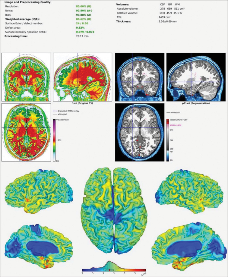
Materials and methods: This cross-sectional study included 222 participants aged 18-60 years, categorized into four groups based on BMI and physical activity levels. Brain MRI scans were acquired using a 3-Tesla scanner. Volumes of various brain regions were calculated and normalized to total intracranial volume. One-way ANOVA and Games-Howell post-hoc tests were used to analyse differences across groups.
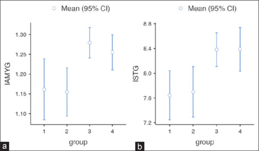
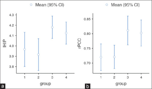
Results: Non-sedentary groups exhibited larger brain volumes in multiple regions compared to sedentary groups, regardless of weight status. This was particularly evident in the posterior cingulate cortex, left hippocampus, and left amygdala. The non-sedentary normal weight group showed significantly larger left middle temporal gyrus volume compared to both sedentary groups. Total intracranial volume was larger in the non-sedentary overweight group compared to the sedentary normal weight group.
Conclusion: Physical activity may have a more pronounced effect on brain volumes than BMI alone, particularly in regions associated with memory and emotion processing. These findings suggest that regular physical activity might confer neuroprotective effects, even in individuals with higher BMI. Public health policies should emphasize increasing physical activity levels across all weight categories to promote both physical and cognitive health.

 求助内容:
求助内容: 应助结果提醒方式:


