{"title":"Computed tomographic appearance of laryngeal lesions in 7 dogs.","authors":"Anna Slusarek, Annick Hamaide, Michaël Lefebvre, Marianne Heimann, Frédéric Billen, Géraldine Bolen","doi":"10.3389/fvets.2025.1633591","DOIUrl":null,"url":null,"abstract":"<p><strong>Objective: </strong>To describe the computed tomographic (CT) features of neoplastic and inflammatory laryngeal masses. The authors hypothesized that specific CT features may help differentiate between these two origins and that regional lymph nodes would be larger in cases of laryngeal neoplasia.</p><p><strong>Methods: </strong>Medical records from two veterinary referral hospitals were screened for dogs diagnosed with either an inflammatory or neoplastic laryngeal mass who underwent CT scans of the neck. Information retrieved from medical records included signalment, physical examination findings, CT scan findings, and definitive diagnosis of the laryngeal mass based on cytological or histopathological results.</p><p><strong>Results: </strong>Four dogs had laboratory reports compatible with a malignant neoplasia and three with an inflammatory process. The shape of the mass was defined as \"ovoid\" in all neoplastic masses and as \"thickening\" in cases of inflammatory processes. Masses were of various sizes (median length: 42 mm, range: 26-82 mm) and either unilateral (1/4 and 2/3 of neoplastic and inflammatory masses respectively) or bilateral. They were described as mineralized (1/4 and 1/3) and as having either an internal (1/4), external (2/4) growth pattern or both (1/4, 3/3). All masses had ill-defined margins and showed heterogeneous contrast enhancement. Two neoplastic and two inflammatory masses had a cavitary aspect. All but one case were associated with regional lymphadenopathy. Thyroid cartilage destruction was observed with two neoplastic and two inflammatory masses.</p><p><strong>Clinical relevance: </strong>This case series describes CT features of laryngeal masses. The shape of the laryngeal mass may assist in determining its nature, inflammatory process was defined as \"thickening\" of the larynx and neoplasia as \"ovoid\"-shaped, whereas other studied features were inconsistently observed in both neoplastic and inflammatory conditions.</p>","PeriodicalId":12772,"journal":{"name":"Frontiers in Veterinary Science","volume":"12 ","pages":"1633591"},"PeriodicalIF":2.9000,"publicationDate":"2025-09-04","publicationTypes":"Journal Article","fieldsOfStudy":null,"isOpenAccess":false,"openAccessPdf":"https://www.ncbi.nlm.nih.gov/pmc/articles/PMC12444889/pdf/","citationCount":"0","resultStr":null,"platform":"Semanticscholar","paperid":null,"PeriodicalName":"Frontiers in Veterinary Science","FirstCategoryId":"97","ListUrlMain":"https://doi.org/10.3389/fvets.2025.1633591","RegionNum":2,"RegionCategory":"农林科学","ArticlePicture":[],"TitleCN":null,"AbstractTextCN":null,"PMCID":null,"EPubDate":"2025/1/1 0:00:00","PubModel":"eCollection","JCR":"Q1","JCRName":"VETERINARY SCIENCES","Score":null,"Total":0}
引用次数: 0
Abstract
Objective: To describe the computed tomographic (CT) features of neoplastic and inflammatory laryngeal masses. The authors hypothesized that specific CT features may help differentiate between these two origins and that regional lymph nodes would be larger in cases of laryngeal neoplasia.
Methods: Medical records from two veterinary referral hospitals were screened for dogs diagnosed with either an inflammatory or neoplastic laryngeal mass who underwent CT scans of the neck. Information retrieved from medical records included signalment, physical examination findings, CT scan findings, and definitive diagnosis of the laryngeal mass based on cytological or histopathological results.
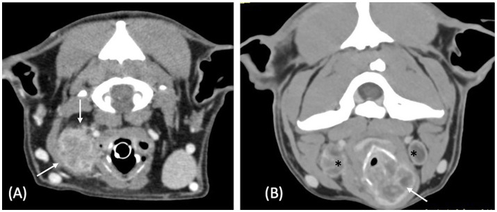
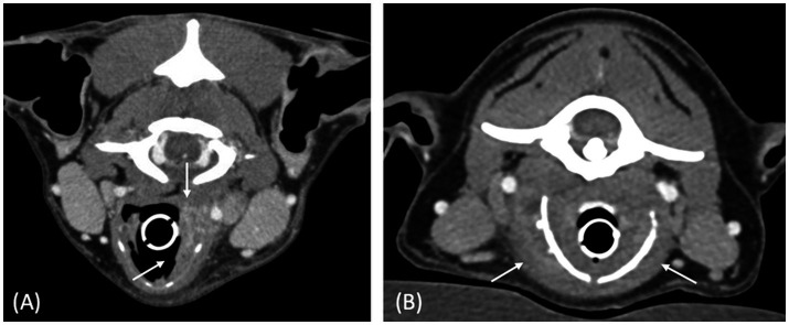
Results: Four dogs had laboratory reports compatible with a malignant neoplasia and three with an inflammatory process. The shape of the mass was defined as "ovoid" in all neoplastic masses and as "thickening" in cases of inflammatory processes. Masses were of various sizes (median length: 42 mm, range: 26-82 mm) and either unilateral (1/4 and 2/3 of neoplastic and inflammatory masses respectively) or bilateral. They were described as mineralized (1/4 and 1/3) and as having either an internal (1/4), external (2/4) growth pattern or both (1/4, 3/3). All masses had ill-defined margins and showed heterogeneous contrast enhancement. Two neoplastic and two inflammatory masses had a cavitary aspect. All but one case were associated with regional lymphadenopathy. Thyroid cartilage destruction was observed with two neoplastic and two inflammatory masses.
Clinical relevance: This case series describes CT features of laryngeal masses. The shape of the laryngeal mass may assist in determining its nature, inflammatory process was defined as "thickening" of the larynx and neoplasia as "ovoid"-shaped, whereas other studied features were inconsistently observed in both neoplastic and inflammatory conditions.
期刊介绍:
Frontiers in Veterinary Science is a global, peer-reviewed, Open Access journal that bridges animal and human health, brings a comparative approach to medical and surgical challenges, and advances innovative biotechnology and therapy.
Veterinary research today is interdisciplinary, collaborative, and socially relevant, transforming how we understand and investigate animal health and disease. Fundamental research in emerging infectious diseases, predictive genomics, stem cell therapy, and translational modelling is grounded within the integrative social context of public and environmental health, wildlife conservation, novel biomarkers, societal well-being, and cutting-edge clinical practice and specialization. Frontiers in Veterinary Science brings a 21st-century approach—networked, collaborative, and Open Access—to communicate this progress and innovation to both the specialist and to the wider audience of readers in the field.
Frontiers in Veterinary Science publishes articles on outstanding discoveries across a wide spectrum of translational, foundational, and clinical research. The journal''s mission is to bring all relevant veterinary sciences together on a single platform with the goal of improving animal and human health.
 求助内容:
求助内容: 应助结果提醒方式:


